Analysis of three-dimensional microarchitecture and degree of mineralization in bone metastases from prostate cancer

Inquiry number
SOL-0000000996
Beamline
BL20B2 (Medical and Imaging I)
Scientific keywords
| A. Sample category | inorganic material, biology, medicine |
|---|---|
| B. Sample category (detail) | biology (in vitro), biomolecule, crystal |
| C. Technique | absorption and its secondary process |
| D. Technique (detail) | |
| E. Particular condition | 3D imaging (cf. CT), room temperature |
| F. Photon energy | X-ray (4-40 keV) |
| G. Target information | local structure, morphology |
Industrial keywords
| level 1---Application area | Pharmaceuticals, others |
|---|---|
| level 2---Target | process analytical technology (PAT) |
| level 3---Target (detail) | organism |
| level 4---Obtainable information | density, structure, molphology |
| level 5---Technique | imaging |
Classification
M60.20 X-ray CT
Body text
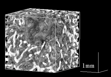
Synchrotron X-ray CT is a nondestructive technique for the precise analysis of bone samples. Using this technique, one can measure three dimensional microarchitecture and the degree of mineralization in cancellous as well as cortical bone. The use of a monoenergetic and parallel synchrotron beam allows precise and quantitative measurements compared with the conventional microfocus X-ray CT.
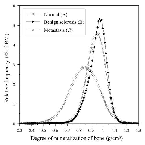
The figure shows three dimensional trabecular architecture and the histogram of degree of mineralization in bone metastasis of prostate cancer. These data reveal the fact that sclerotic site of bone metastasis of prostate cancer is mixed with areas of bone resorption, and shows the different feature of bone mineralization compared as other osteosclerotic lesions.
[ T. Sone, T. Tamada, Y. Jo, H. Miyoshi and M. Fukunaga, Bone 315, 432-438 (2002), Fig. 3A, 4,
©2002 Elsevier, Inc. ]
Source of the figure
Original paper/Journal article
Journal title
Bone. 2004 Aug;35(2):432-8.
Figure No.
Technique
Source of the figure
No figure
Required time for experimental setup
3 shift(s)
Instruments
| Instrument | Purpose | Performance |
|---|---|---|
| X-ray CT system | Obtain the information of 3-D internal structer | spatial resolution of about 10um |
References
Related experimental techniques
Questionnaire
This solution is an application of a main instrument of the beamline.
Ease of measurement
Easy
Ease of analysis
Middle
How many shifts were needed for taking whole data in the figure?
Two-three shifts