Successful Demonstration of High-Resolution X-ray Imaging of Thick Objects (Press Release)
- Release Date
- 03 Feb, 2014
- BL29XU (RIKEN Coherent X-ray Optics)
Osaka University
RIKEN
Key points
• World’s first demonstration of X-ray ptychography using multislice approach
• Expected application to nondestructive high-resolution three-dimensional observation of integrated circuits and biological bone tissue
|
A research group successfully demonstrated that high-resolution X-ray imaging can be applied to thick objects, which previously was impossible by conventional methods. The group was led by Yukio Takahashi (associate professor) and Akihiro Suzuki (1st year Ph.D. student, fellow of Japan Society for the Promotion of Science) of the Graduate School of Engineering, Osaka University, and Tetsuya Ishikawa (Director) of RIKEN SPring-8 Center. X-rays exhibit high transmittance and can be used to observe the internal structure of even thick objects. They are electromagnetic waves with a wavelength on the angstrom (Å) order (1 Å is one-ten-billionth of a meter). Hence, the development of a high-resolution X-ray microscope is possible in principle. However, the high-resolution observation of the inside of thick objects is not easy. In X-ray imaging, projection approximation is generally adopted, in which a specimen is projected in the direction of X-ray propagation and approximated to a very thin object. A thick object, however, cannot be observed at a high resolution by this method because X-ray wavefronts change in the specimen. To address this issue, the research group adopted a multislice approach*1 and demonstrated the validity of X-ray ptychography*2 by this approach for the first time in the world. In the multislice approach, a specimen is modeled as an accumulation of thin layers perpendicular to incident X-rays, and the changes in X-ray wavefronts between the layers are measured. Multislice X-ray ptychography performed at SPring-8*3 revealed that a resolution of ~50 nm (1 nm is one-billionth of a meter), much better than the limit under the projection approximation (192 nm), was achieved for a specimen with a thickness of 105 μm (1 μm is one-millionth of a meter). The demonstrated multislice X-ray ptychography enables the observation of nanostructures and tissues in thick objects. It can be applied to the nondestructive high-resolution three-dimensional observation of, for example, the nanowiring of integrated circuits and biological bone tissue. The achievements of this research were published online in the American scientific journal Physical Review Letters on 4 February 2014. Publication: |
<<Figures>>
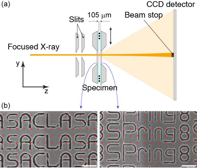
(b) Electron microscopy images of specimen.
7 keV synchrotron radiation X-rays are focused on an ~500-nm-diameter spot through total reflection mirrors. A double-layered specimen with a gap of 105 μm is placed on the focal point and the intensity of X-rays scattered at the specimen is measured using a charge-coupled device (CCD) detector.
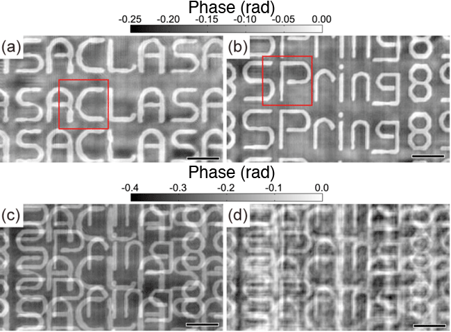
reconstructed by multislice approach. (c) Projected image obtained by superimposing
maps (a) and (b). (d) Projected image reconstructed by conventional method.
Images of the first and second layers of a double-layered specimen are independently reconstructed by the multislice approach. The spatial resolution is low and many artifacts are observed in the image obtained by the conventional method, whereas the spatial resolution is high and few artifacts are observed in the images obtained by the multislice approach.
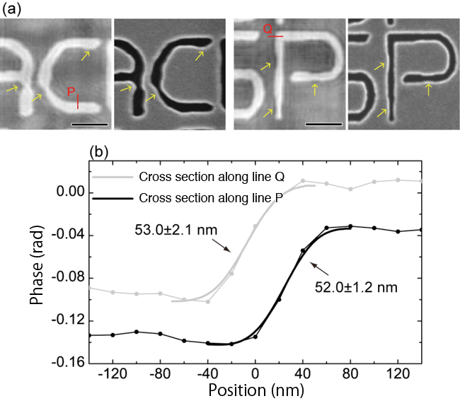
2(a) and 2(b). (a, right) Electron microscopy images of the same regions.
(b) Cross-sectional profiles of positions indicated by red lines.
Fine structures are in agreement between the phase maps reconstructed by the multislice approach and the electron microscopy images, showing the high spatial resolution of this approach. Cross-sectional profiles reveal that the spatial resolution is ~50 nm.
<<Glossary>>
*1 Multislice approach
A wave-optics analysis method in which a specimen is modeled as an accumulation of thin layers perpendicular to the incident light and the light is assumed to travel through the layers in turn.
*2 X-ray ptychography
A type of coherent X-ray diffraction imaging. By scanning a specimen two-dimensionally, coherent diffraction patterns are taken at several different points in overlapping X-ray irradiation regions. A phase retrieval algorithm is applied to the diffraction patterns to reconstruct an image of the specimen. “Ptycho” means “fold” in Greek (πτνξ).
*3 SPring-8
SPring-8 is a shared synchrotron radiation facility that delivers the world’s highest-brilliance synchrotron radiation. It is owned by RIKEN and located in Harima Science Park City, Hyogo Prefecture, Japan. The name “SPring-8” is derived from “Super Photon ring-8 GeV”. Synchrotron radiation is a type of light radiated when charged particles are forced to bend in magnetic fields. SPring-8 can produce X-rays with a high coherence because of the small size of circulating electron groups and high stability.
|
For more information, please contact: |

- Previous Article
- Successful Synthesis of Artificial Rhodium (Press Release)
- Current article
- Successful Demonstration of High-Resolution X-ray Imaging of Thick Objects (Press Release)