Snapshot Measurement of "Color" of XFEL - Important step for developing XFEL science - (Press Release)
- Release Date
- 28 Aug, 2006
- XFEL
Key Points:
• Development of spectrometer for XFEL diagnostics
• First realization of high-resolution, single-shot measurement
• Success of international collaboration between major XFEL projects
|
The research group of RIKEN (President Ryoji NOYORI), the Japan Synchrotron Radiation Research Institute (JASRI, Director General Akira KIRA), and the Stanford Linear Accelerator Center (SLAC, Director Jonathan Dorfan) has announced the development of a new instrument for measuring the energy spectrum of an X-ray free-electron laser (XFEL). This development was achieved by Dr. Makina Yabashi and Dr. Tetsuya Ishikawa (Group Director) of the "RIKEN-JASRI Joint-Project for the SPring-8 XFEL" (Director Toichi SAKATA) and Dr. Jerome Hastings (Project Director) at SLAC, collaborating with groups at Osaka University and the Lawrence-Berkeley National Laboratory. Article: |
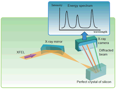 Fig. 1. Principle of spectrometer
The incident beam, which is focused and diverged by the X-ray mirror,
is diffracted by the perfect crystal of silicon.
The spatial profile of the diffracted beam is recorded using the X-ray camera.
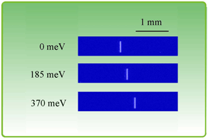 Fig. 2. Profile of diffracted beam
The position of the diffracted beam is shifted as the incident wavelength (photon energy) changes.
The numbers on the left indicate deviations of the incident photon energy.
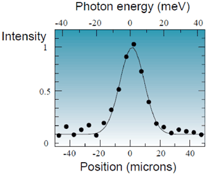 Fig. 3. Energy resolution
Measurement result of energy resolution using monochromatic X-rays as incident beam.
A resolution of 13.1 meV was obtained by subtracting the energy spread of the incident beam.
For more information, please contact:
Dr. Makina Yabashi,
RIKEN-JASRI Joint-Project for the SPring-8 XFEL
Phone +81 -(0)791-58-0831
Fax +81 -(0)791-58-0830
e-mail: yabashi@spring8.or.jp
- Previous Article
- Phantom of bulk metallic glass Zr and Ti (Press Release)
- Current article
- Snapshot Measurement of "Color" of XFEL - Important step for developing XFEL science - (Press Release)