Discovery of Inhomogeneous Microstructure of Liquid Water Previously Considered to Have Uniform Density - Clarification of Mystery of Transparent Water by Observation Using Synchrotron Radiation in Japan and the US (Press Release)
- Release Date
- 11 Aug, 2009
- BL17SU (RIKEN Coherent Soft X-ray Spectroscopy)
RIKEN
SLAC* National Accelerator Laboratory
Stockholm University
Key research findings
• The inhomogeneity of water is caused by the coexistence of two types of microstructure in water.
• The size of one type of inhomogeneous microstructure, similar to ice, is approximately 1 nm in diameter.
• Clarification of the temperature dependence of the microstructures will lead to an understanding of the role of water in organisms and in chemical reactions.
|
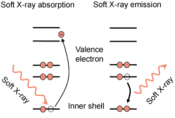
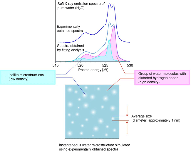
RIKEN (Ryoji Noyori, President) discovered the inhomogeneous state of liquid-water molecules, whose density had previously been considered to be uniform, by observing water microscopically, using two synchrotron radiation facilities, SPring-8*1 in Japan and the Stanford Synchrotron Radiation Lightsource (SSRL)*1 in the US. This discovery was achieved by a joint research group*2 led by Shik Shin, team leader of the Excitation Order Research Team, Quantum Order Research Group at RIKEN SPring-8 Center (Tetsuya Ishikawa, Director) as well as a professor at The Institute for Solid State Physics of The University of Tokyo; Osamu Takahashi, an assistant professor at the Faculty of Science of Hiroshima University; and Anders Nilsson, a professor at the SLAC National Accelerator Laboratory. The inhomogeneity of water density is caused by the coexistence of two types of microstructure in water, which were discovered in 2008. The observation of electron states using the RIKEN Coherent Soft X-ray Spectroscopy Beamline (BL17SU) at SPring-8 revealed that the inhomogeneity appears because icelike microstructures are scattered just like a polka-dot pattern among groups of water molecules with distorted hydrogen bonds. It was also revealed that the size of the icelike microstructures is approximately 1 nm in diameter (1 nm is 10-9 m). Although water has been considered to have uniform density, finding inhomogeneous microstructures in water and clarifying their change with temperature will significantly advance the understanding of the role of water in various phenomena such as in organisms and in chemical reactions as well as the mechanism underlying the dissolution of substances in water. The research achievements were published in the online version of the US academic journal Proceedings of the National Academy of Sciences on 13 August 2009. Publication: |
* SLAC: Stanford Linear Accelerator Center
<Figure>
|
|
||||||
|
|
||||||
<Glossary>
*1 SPring-8, a large synchrotron radiation facility, and Stanford Synchrotron Radiation Lightsource (SSRL)
SPring-8 is a RIKEN facility located at Harima Science Garden City, Hyogo Prefecture, that can provide the most powerful synchrotron radiation available to date. The name SPring-8 is derived from Super-Photon ring-8 GeV. Synchrotron radiation is a narrow, powerful beam of electromagnetic radiation generated when electron beams, accelerated to nearly the speed of light, are forced to travel in a curved path by an electromagnet. Studies conducted at SPring-8 using synchrotron radiation include those on nanotechnology, biotechnology, and industrial applications. SSRL is a synchrotron radiation facility established in the SLAC National Accelerator Laboratory (SLAC) in the US.
*2 Joint research group
The joint research team mainly consisted of Shik Shin, team leader, Takashi Tokushima, a research scientist, Yuka Horikawa, a junior research associate, and Yoshihisa Harada, a visiting scientist (also a research associate professor at the School of Engineering of The University of Tokyo) of the Excitation Order Research Team, Quantum Order Research Group at RIKEN SPring-8 Center; Osamu Takahashi, an assistant professor at the Faculty of Science of Hiroshima University; Anders Nilsson, a professor at the SLAC National Accelerator Laboratory; and L. G. M. Pettersson, a professor at Stockholm University in Sweden.
|
For more information, please contact: Dr. Takashi Tokushima (RIKEN) or for RIKEN Coherent Soft X-ray Spectroscopy Beamline (BL17SU), |
 ,
, ,
, ,
, .
.- Previous Article
- Dramatically Breaking World Record for Strength of Magnetic Field in Ultrahigh-Magnetic-Field X-Ray Spectroscopic Experiment (Press Release)
- Current article
- Discovery of Inhomogeneous Microstructure of Liquid Water Previously Considered to Have Uniform Density - Clarification of Mystery of Transparent Water by Observation Using Synchrotron Radiation in Japan and the US (Press Release)