Success in Determining Structure of a Single Catalyst Particle That Produces Hydrogen from Methane Using X-ray μ-Beam
- Release Date
- 08 Jun, 2011
- BL37XU (Trace Element Analysis)
Institute for Molecular Science,
National Institutes of Natural Sciences
Japan Synchrotron Radiation Research Institute
|
A research group led by Mizuki Tada, an associate professor of the Institute for Molecular Science, National Institutes of Natural Sciences, and Tomoya Uruga, an associate chief scientist of the Japan Synchrotron Radiation Research Institute (JASRI), has succeeded in determining the structure of a single catalyst particle that produces hydrogen from methane using an X-ray microbeam (μ-beam) at SPring-8, one of the world’s leading synchrotron radiation facilities. Previously, the structure of solid catalyst particles, which play various roles in chemical reactions, was only observed as assemblies or mixtures of particles with different structures and electron states. It is very difficult to determine the detailed features and catalytic functions of solid catalysts from averaged information obtained from the assemblies and mixtures of nonuniform particles. In this research, scientists in the group produced an X-ray μ-beam with a width of 1000 nm (a nanometer is one-billionth of a meter) and a height of 800 nm at SPring-8, and attempted to clarify the structure of a single catalyst particle with a size equivalent to that of the X-ray μ-beam. As a result, they succeeded in clarifying, for the first time in the world, the local coordination structure*2 of a supported Ni catalyst,*1 which produces hydrogen from methane, by the X-ray absorption fine structure (XAFS)*3 method at a spatial resolution equivalent to the size of the single catalyst particle. This analytical technique can be applied to the structural analysis of various catalyst particles. In the future, this technique is expected to lead to the clarification of the principle of the synergetic effect of assemblies and mixtures of particles with different properties, shapes, and sizes and to the development of more efficient catalysts. The results of this study were published online in the physics and chemistry journal of the Royal Society of Chemistry (UK) Physical Chemistry Chemical Physics on 8 June 2011. The research achievements were highly acclaimed and highlighted on the cover page of the corresponding issue. Publication: |
<<Glossary>>
*1 Supported Ni catalyst
Supported Ni catalysts are catalysts in which Ni particles are placed on an oxide support. In this study, a catalyst in which Ni particles are dispersed and supported on the surface of an oxide solid solution Ce2Zr2Oy was used.
*2 Local coordination structure
A local coordination structure refers to a structure very near the element to be measured. This provides information on the type and number of atoms around the element (coordination number) and on the distance between the atoms (bond length). The local coordination structure can be clarified by analyzing extended X-ray absorption fine structure (EXAFS) spectra.
*3 X-ray absorption fine structure (XAFS)
When synchrotron radiation X-rays with a particular energy are irradiated onto a material, an X-ray absorption spectrum can be obtained. The symmetry and the valence for the element to be measured can be determined by analysis in the vicinity of the absorption edges in this spectrum, i.e., the X-ray absorption near edge structure (XANES) spectrum. Moreover, the type and number of atoms around the element to be measured and the distance between the atoms, i.e., local coordination structures, can be determined by analyzing EXAFS spectra. The XAFS method is almost the only technique that can reveal the structure of materials without long-range structural order, such as catalyst particles. The XAFS method with hard X-rays enables the in situ structural analysis under catalytic reaction conditions.
<<Figures>>
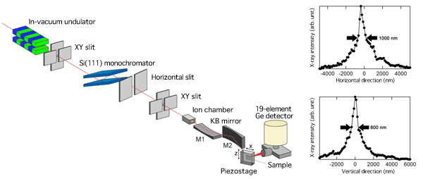
and beam size profile of X-ray μ-beam at 8 keV (right)
The X-ray μ-beam is concentrated in both the horizontal (width) and vertical (height) directions.
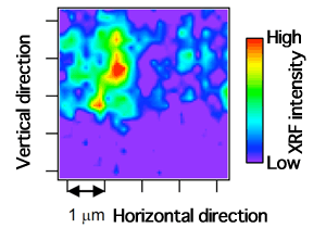
The position of a catalyst particle on a thin-film substrate can be specified because the X-ray intensity increases at positions where the catalyst particle is present.
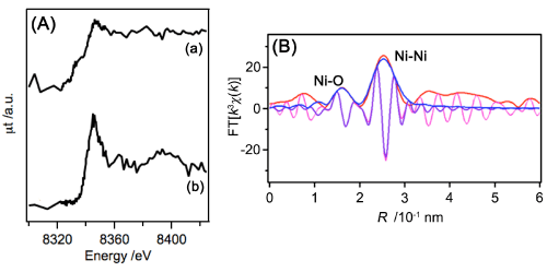
(A) μ-XANES spectra for a single catalyst particle
(a) Active Ni catalyst obtained by reduction
(b) Inactive Ni catalyst obtained by oxidation
(B) Fourier transformation of μ-EXAFS spectra for a single catalyst particle and curve-fitting analysis
|
For more information, please contact: |


- Previous Article
- First X-ray lasing of SACLA (Press Release)
- Current article
- Success in Determining Structure of a Single Catalyst Particle That Produces Hydrogen from Methane Using X-ray μ-Beam