Development of Novel Method for Utilizing Coherent X-rays (Press Release)
- Release Date
- 07 Mar, 2013
- BL29XU (RIKEN Coherent X-ray Optics)
Osaka University
RIKEN
Key research findings
• Visualization of nanoscale strain distribution in a thick sample using Bragg diffraction of coherent X-rays
• Proposed new method for producing an X-ray vortex microbeam using the phase singularities of a strain field
|
A research group led by Yukio Takahashi (associate professor) at the Graduate School of Engineering, Osaka University, and Tetsuya Ishikawa (chief scientist) at RIKEN SPring-8 Center, visualized the dislocation strain fields in a sample and developed a novel method for utilizing coherent X-rays to produce an X-ray vortex beam. Dislocations refer to linear defects in crystals, which induce local strain around the dislocations. Dislocation strain fields play a significant role in the mechanical and electrical conduction properties of materials. Dislocations are generally observed by X-ray topography and transmission electron microscopy, which are limited in terms of spatial resolution and measurable thickness. The scientists in the research group developed a method called Bragg X-ray ptychography using the RIKEN Physics Beamline I (BL29XUL) at SPring-8.*1 They succeeded in visualizing the strain fields caused by dislocations in a 1-μm-thick silicon single crystal with nanometer-order spatial resolution. This visualization technique is expected to be used in the design and development of new structural materials and semiconductor materials. The research group also found that X-rays with a spiral wavefront (i.e., an X-ray vortex beam) can be formed using the strain fields caused by dislocations in the silicon single crystal. X-ray vortex beams have an important characteristic that the mode of orbital angular momentum*2 can be controlled by selecting the phase singularities and reflecting surface. This is a very interesting finding from the viewpoint of X-ray optics because the formation of vortex beams using X-rays has seldom been reported, although their formation using visible light and electron beams has been frequently reported. X-ray vortex beams are considered to exhibit strong dichroism and are expected to be applied to the analysis of complicated electronic structures in materials. This research was supported by a Grant-in-Aid for Challenging Exploratory Research of Japan Society for the Promotion of Science under the title of Demonstration of Atomic-Resolution X-ray Microscopy and Its Application to Dislocation Imaging (Representative, Yukio Takahashi). The results were published online in the journal of the American Physical Society Physical Review B Rapid Communications on 7 March 2013. Publication: |
<<Figures>>
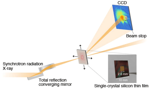
Synchrotron radiation X-rays of 11.8 keV are focused onto an approximately 1-μm-diameter spot using total-reflection converging mirrors. A single-crystal silicon thin film is placed on the focal point, and the coherent Bragg diffraction pattern from the (220) crystal plane is measured using a charge-coupled device (CCD).
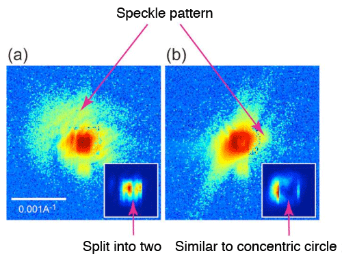
in single-crystal silicon thin film (shown with logarithmic scale)
The centers of the patterns are shown with a linear scale in the right lower insets. (a) When the thin film is irradiated with X-rays crossing the dislocation. (b) When the dislocation core is irradiated with X-rays.
When the single-crystal silicon thin film was irradiated with coherent X-rays, a characteristic coherent Bragg diffraction pattern was observed. When X-rays were introduced in the direction crossing the dislocation, the Bragg spot was split into two because of the destructive interference of X-rays, producing a characteristic speckle pattern. When the dislocation core is irradiated with X-rays, the Bragg spot forms a shape similar to a concentric circle.
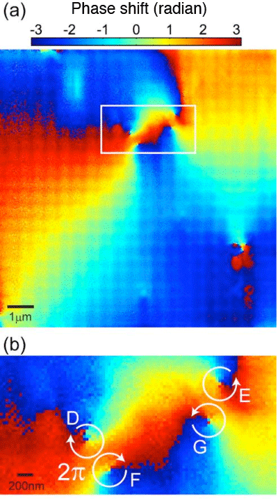
by Bragg X-ray ptychography. (b) Magnified view of the boxed region in (a).
The phase sharply changes in the region distorted by dislocations. There are four phase singularities, around which the phase changes in a spiral manner.
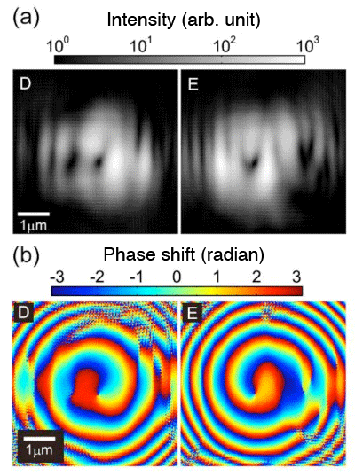
by wave optics simulation. (Phase singularities D and E in Fig. 3 were
irradiated with X-rays and the wave field of the diffraction wave
transmitted 10 mm from the sample was calculated.)
The simulation results indicate that an X-ray vortex beam can be formed by irradiating the phase singularities with focused X-ray beams. In other words, the mode of the orbital angular momentum can be changed by selecting the phase singularities.
<<Glossary>>
*1 SPring-8
SPring-8 is a shared synchrotron radiation facility that delivers the world’s highest-brilliance synchrotron radiation. It is owned by RIKEN and located in Harima Science Park City, Hyogo Prefecture, Japan. The name “SPring-8” is derived from “Super Photon ring-8 GeV”. Synchrotron radiation is a type of light radiated when charged particles are forced to bend in magnetic fields. SPring-8 can produce X-rays with a high coherence because of the small size of circulating electron groups and high stability.
*2 Orbital angular momentum
The orbital angular momentum is the cross product of the position coordinate and conjugate momentum and can be used to determine the quantum number (an index for the quantum state) of electrons, which are bound to central force fields such as those of atoms.
|
For more information, please contact: |

- Previous Article
- X-ray observation of a helium atom and placing a nitrogen atom inside He@C60 and He@C70 (Press Release)
- Current article
- Development of Novel Method for Utilizing Coherent X-rays (Press Release)