Development of Highly Efficient X-ray Absorption Spectroscopy (XAS) Using Characteristics of X-ray Free Electron Laser (XFEL) Pulses (Press Release)
- Release Date
- 24 Sep, 2013
- SACLA
RIKEN
Japan Synchrotron Radiation Research Institute (JASRI)
Kyoto University
Tokyo University of Agriculture and Technology
Key points
• Successful demonstration of new XAS at SACLA*1, RIKEN’s XFEL facility
• Simultaneous monitoring of broad X-ray absorption spectra using two X-ray beams
• A step toward establishing a technique for tracking the motion of atoms and molecules during ultrafast chemical reactions
|
A joint research group comprising RIKEN (President, Ryoji Noyori), JASRI (President, Yoshiharu Doi), Kyoto University (President, Hiroshi Matsumoto), and Tokyo University of Agriculture and Technology (President, Tadashi Matsunaga) has developed a new technique of X-ray absorption spectroscopy*2 using an X-ray free electron laser (XFEL)*1 and carried out a successful demonstrative test at SACLA, an XFEL facility of RIKEN. The group was led by Tetsuo Katayama (research scientist) from JASRI; Makina Yabashi (group director) from RIKEN SPring-8 Center (Director, Tetsuya Ishikawa); Yoshihiro Ogi (senior research scientist) from the RIKEN Center for Advanced Photonics (Director, Katsumi Midorikawa); Toshinori Suzuki (professor) from the Graduate School of Science, Kyoto University; and Kazuhiko Misawa (professor) from the Graduate School of Engineering, Tokyo University of Agriculture and Technology. The distribution of electrons in atoms and molecules and the spatial geometric structure of atomic nuclei are closely related to the chemical responsiveness of the atoms and molecules. X-ray absorption spectroscopy is one of the effective techniques for observing the electronic and geometric structures of substances. For example, irradiating substances with X-ray pulses for a time as short as the duration of a chemical reaction will enable us to observe and track the chemical reaction in real time, leading to the clarification of the entire mechanism of the reaction. However, the time width*3 of synchrotron X-ray pulses widely used in conventional X-ray absorption spectroscopy is several tens of picoseconds (1 picosecond = one-trillionth of a second), which is too long to track ultrafast chemical reactions that occur in about one femtosecond (1 femtosecond = one-quadrillionth of a second). XFEL is a light source that can generate X-ray pulses with a time width of about 10 fs, 104-fold shorter than that of synchrotron X-ray pulses, and is highly promising for use in the real-time observation of chemical reactions. Compared with synchrotron light sources, however, the current SACLA generates only 20 X-ray pulses per second and is inferior in terms of the efficiency of time-scale measurements. Therefore, the development of a highly efficient spectroscopy method that can provide much more information per pulse has been required to fully use the XFEL. The joint research group developed a new technique for simultaneously measuring the dependence of X-ray absorbance on photon energy (i.e., obtaining X-ray absorption spectra) using a single X-ray pulse with the energy range of XFEL pulses. To simultaneously obtain X-ray absorbance spectra with a single pulse, X-ray spectra before and after X-ray irradiation must be simultaneously obtained. In the developed technique, the X-ray from the XFEL is split into two beams using a transmission diffraction grating*4 and the absorbances of the reference and transmitted X-rays are simultaneously measured for each pulse to obtain absorption spectra. The absorption spectra obtained by this technique were in good agreement with those obtained by conventional measurement methods, demonstrating the effectiveness of the technique. The developed technique is expected to be widely used in X-ray absorption spectroscopy using XFEL for time-resolved measurements, such as in the real-time observation of chemical reactions. The achievements of this research were published online in the American scientific journal Applied Physics Letters on 25 September 2013. This research was supported by the XFEL Intensive Strategy Project, the Ministry of Education, Culture, Sports, Science and Technology. |
<<Figures>>
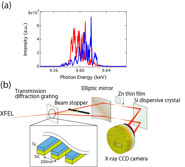
(b) X-ray beams split using transmission diffraction grating and spectrometer
consisting of elliptic mirror, Si dispersive crystal, and X-ray charge-coupled
device (CCD) camera
(a) Spectrum of SASE XFEL. Red and blue spectra are obtained with a single pulse. The spectra exhibit a random spike pattern.
(b) Diffracted light from a transmission diffraction grating (Ta, tantalum; SiC, silicon carbide) was used to simultaneously obtain two types of spectra. The divergence angle (the degree of expansion of the beam) of X-ray beams was increased using an elliptic mirror to reflect the beams because a large divergence angle is required to obtain broad energy spectra. When two split beams are incident to a Si dispersive crystal, they are diffracted at different angles in accordance with the X-ray energy to satisfy the Bragg condition (the diffraction angle of X-rays depends on the X-ray energy). The reflected beams were detected using an X-ray CCD camera.
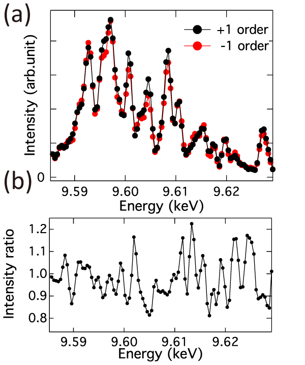
(a) Spectra obtained by two diffracted light beams without sample.
(b) Ratio of intensity indicated by the black line to that indicated by the red line in (a). The two spectra are in good agreement, indicating the possibility of normalization to determine absorption spectra.
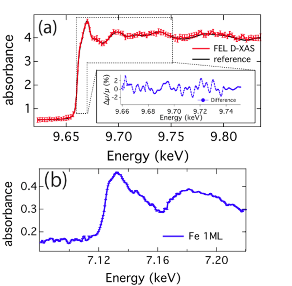
X-ray absorption spectra obtained by placing different samples on one of the two light paths. The samples used were (a) Zn thin film (indicated by red line) and (b) aqueous solution of ferric ammonium complex. The black line in Fig. 3(a) shows the reference X-ray absorption spectrum obtained by the conventional method with synchrotron radiation.
<<Glossary>>
*1 X-ray free electron laser (XFEL) and SACLA
XFEL refers to a laser with wavelengths in the X-ray region. Unlike conventional lasers that oscillate using semiconductors or gas as a medium, an XFEL uses electron beams that rapidly travel in vacuum as a medium and hence, in principle, has no wavelength limit. SACLA is Japan’s first XFEL and was constructed jointly by RIKEN and JASRI. SACLA is one of the five national core technologies in the Science and Technology Basic Plan. The construction and preparation of SACLA was launched in FY2006 in a five-year project and was completed in March 2011. The name SACLA is short for SPring-8 angstrom compact free electron laser. In June 2011, the first oscillation of the X-ray laser was achieved. Since March 2012, SACLA has been open to public users who wish to carry out their own experiments. Despite it being several times smaller than similar overseas facilities, SACLA is capable of generating lasers with the world’s shortest wavelength of ≤0.1 nm.
*2 X-ray absorption spectroscopy (XAS)
When a sample is irradiated with X-rays, those with an energy specific to the constituent elements are absorbed by the sample. XAS is an experimental technique for measuring the absorbance of samples by varying the energy of X-rays used for irradiation and can provide information on the local structure and chemical state in the vicinity of atoms of interest. XAS can be applied to various samples because they need not be crystalline and, with the exception of light elements, can be measured in air.
*3 Time width of pulse (pulse width)
Pulses are generated only for a certain duration (time width). The period of pulses is equal to the number of pulses generated in one second and is not directly related to the time width of pulses.
*4 Transmission diffraction grating
An optical element with a periodic concave-convex structure. When a transmission diffraction grating is irradiated with X-rays, the light transmitted through the grating and that scattered at the grating interfere with each other, causing intense diffracted waves (diffracted light) to propagate in a certain direction.
|
For more information, please contact: |
- Current article
- Development of Highly Efficient X-ray Absorption Spectroscopy (XAS) Using Characteristics of X-ray Free Electron Laser (XFEL) Pulses (Press Release)