BL20XU OUTLINE
Inquiry number
INS-0000000286
ABSTRACT
The beamline 20XU is designed for application to various imaging technologies: X-ray microscopy, micro-tomography, refraction-enhanced imaging, etc. The BL20XU is the second medium-length beamline in the SPring-8.
AREA OF RESEARCH
- X-ray micro-/nano-imaging: micro-CT, nano-CT (15-37.7 keV), refraction/phase contrast imaging, X-ray diffraction tomography (XRD-CT, DCT), microbeam/scanning x-ray microscope
- Research and development of X-ray optics and optical elements, coherent X-ray optic
- Ultra small-angle X-ray scattering (USAXS, 23 keV)
KEYWORDS
- Scientific field
micro-imaging, coherent optics, ultra-small-angle scattering - Equipment
- High resolution X-ray imaging detectors (CCD camera coupled with optical lens and phosphor screen (beam monitor type II and type III
- Beam monitor type II: field of view 3-6 mm, pixel size 0.3-0.6 µm
Beam monitor type III: field of view 0.5-2 mm, pixel size 0.5-2 µm - X-ray Image intensifier (Be window, 4 inches type)
- High-energy X-ray phase-contrast full-field tomographic microscopy
- XRD-CT
- DCT
- High resolution X-ray imaging detectors (CCD camera coupled with optical lens and phosphor screen (beam monitor type II and type III
SOURCE AND OPTICS
The light source is hybrid-type "in-vacuum" planar undulator whose periodic length is 26 mm. Maximum K-value is designed to be 2.0 at an undulator magnet gap of 7 mm in order to cover the full energy regions above 7.62 keV. Monochromator is "SPring-8 standard" double crystal monochromator placed at 46 m from the source point. The liquid nitrogen cooling system developed at the BL47XU will be employed. The first crystal of monochromator is combination of Si (111) orientation crystal and Si (220) orientation. These two crystals are interchangeable by using linear-translation stage without venting the monochromator vacuum.
The second crystal is Si 111-orientation. Therefore, by switching only the first crystal, both (+ -) configuration of Si 111-111 and Si 220-220 configuration can be used. Therefore, the energy region from 7.62 to 61 keV is available by the combination of the two reflections.
- Constant exit double crystal monochromator
- SPring-8 standard, cryogenic cooling
- Lattice plane: Si 111 (7.62 - 37.7 keV), Si 220 ( - 61 keV)
- Range of Bragg angle: 3 -15 degree
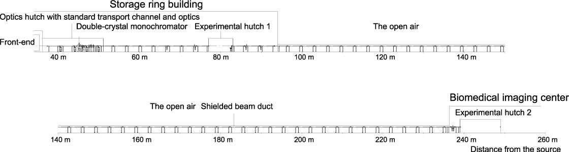
Schematic View of Beamline
- X-rays at Sample
- X-ray beam cross-section: 0.7 mm (vertical) x 1.4 mm (horizontal) at the experimental hutch I (~ 80 m from source point)
- Flux density at the hutch I is about 1013 photons/mm2 with Si 111 refrection
- X-ray beam cross-section at the experimental hutch II (245 m from souce point): ~ 2 mm × 4 mm
- X-ray beam cross-section: 0.7 mm (vertical) x 1.4 mm (horizontal) at the experimental hutch I (~ 80 m from source point)
EXPERIMENTAL STATIONS
Monochromatic beam is extracted into the Medium-length Beamline Facility that is located at about 200 m from storage ring. Two experimental hutches are located at 80 m from source and at 245 m from the source, respectively. The first experimental hutch is in the experimental hall of the storage ring building, and the second one is constructed in the Medium-length Beamline Facility. The first medium length beamline 20B2 is bending-magnet light source beamline and is now used for microscopy, and X-ray topography. The role of BL20XU is complementary to BL20B2. By using high-flux density X-ray beam from the undulator light source, although the beam cross-section is only a few mm even at the end station that is located at 245 m from the undulator, real time observation of live specimen will become possible. It is also to be noticed that a highly coherent X-ray beam can be provided by utilizing the long source to sample distance of 245 m.
- Detectors : conventional type ionization chambers, NaI scintillation counters, pure Ge detector.
- X-ray imaging detectors : CCD camera coupled with optical lens and phosphor screen (beam monitor type II and type III), X-ray zooming tube, X-ray image intensifier, and direct sensing X-ray pickup-tube camera
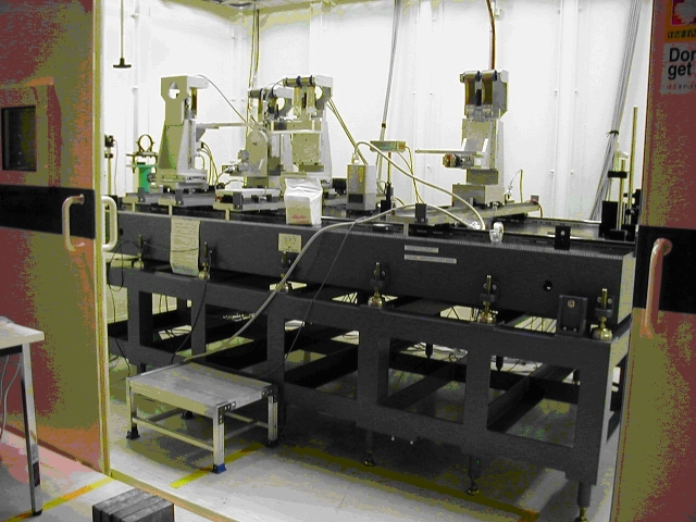
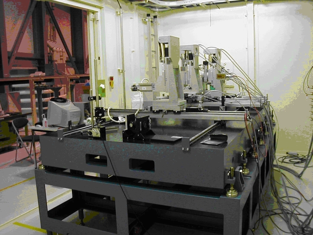
- Versatile high-precision diffractometer for X-ray microscopy at the experimental hutch I
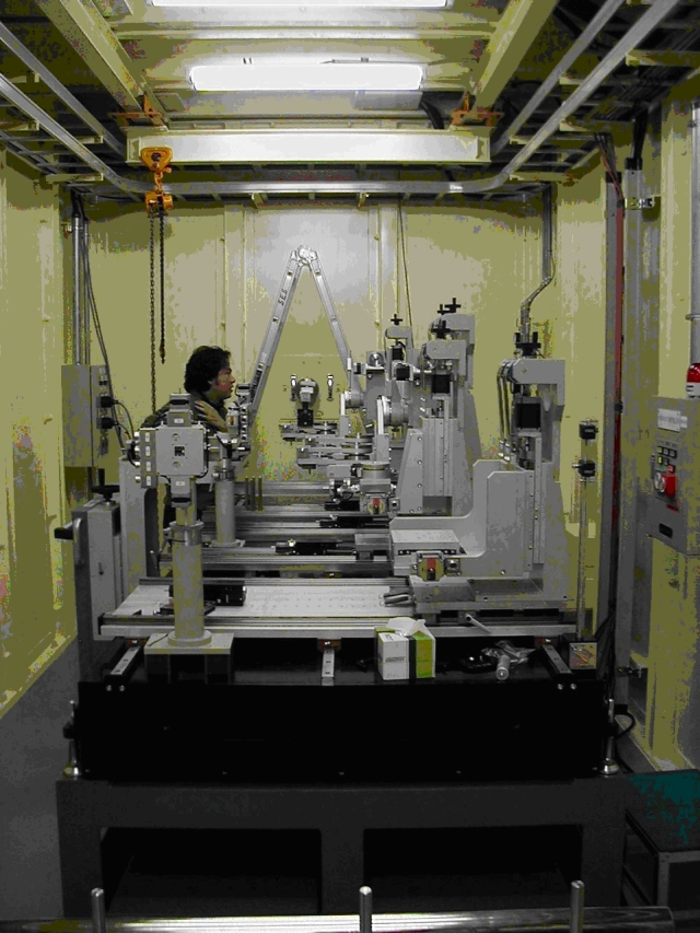
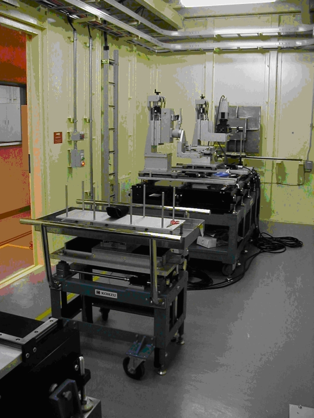
- Multi-purpose high-precision diffractometer for various imaging experiments at the experimental hutch II
PUBLICATION SEARCH
* Sorry, Some parts of results are displayed using Japanese characters.
CONTACT INFORMATION
Please note that each e-mail address is followed by "@spring8.or.jp."
Akihisa TAKEUCHI
SPring-8 / JASRI
1-1-1 Kouto, Sayo-cho, Sayo-gun, Hyogo 679-5198
Phone : +81-(0)791-58-0833
Fax : +81-(0)791-58-0830
e-mail : take
Masayuki UESUGI
SPring-8 / JASRI
1-1-1 Kouto, Sayo-cho, Sayo-gun, Hyogo 679-5198
Phone : +81-(0)791-58-0833
Fax : +81-(0)791-58-0830
e-mail : uesugi
Yuki SADA
SPring-8 / JASRI
1-1-1 Kouto, Sayo-cho, Sayo-gun, Hyogo 679-5198
Phone : +81-(0)791-58-0833
Fax : +81-(0)791-58-0830
e-mail : yuki.sada