X-ray diffraction microscopy
Body text
X-ray diffraction microscopy is a unique technique to reconstruct a sample image from its oversampled diffraction intensity pattern. Using this technique, one can determine electron density distribution without need of sample crystallization.
The following figures show the diffraction intensity pattern and the 2D reconstruction of Escherichia coli bacteria.
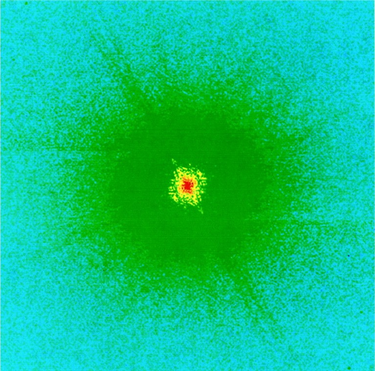
Fig. X-ray diffraction intensity pattern of Escherichia coli bacteria
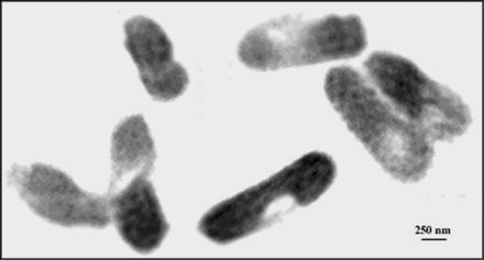
Fig. Image of Escherichia coli bacteria reconstructed from the diffraction intensity pattern
[ J. Miao, K. O. Hodgson, T. Ishikawa, C. A. Larabell, M. A. LeGros and Y. Nishino, Proceedings of National Academy of Science of the USA 100, 110-112 (2003), Fig. 1A, 2,
©2003 National Academy of Science ]
Scientific keywords
| A. Sample category | inorganic material, organic material, biology, medicine |
|---|---|
| B. Sample category (detail) | metal, alloy, semiconductor, insulator, ceramics, solid-state crystal, amorphous, glass, organism, cell, biological material |
| C. Technique | X-ray diffraction |
| D. Technique (detail) | coherent scattering, phase measurement, speckle |
| E. Particular condition | 2D imaging, 3D imaging (cf. CT), X-ray microscopy |
| F. Photon energy | X-ray (4-40 keV) |
| G. Target information | structure analysis |
Industrial keywords
| level 1---Application area | Pharmaceuticals |
|---|---|
| level 2---Target | drug design, food, Environmental material |
| level 3---Target (detail) | organism |
| level 4---Obtainable information | |
| level 5---Technique | diffraction, imaging |
Inquiry number
SOL-0000000970