BL37XU OUTLINE
Inquiry number
INS-0000000591
ABSTRACT
BL37XU is a hard X-ray undulator beamline that is mainly used for studies of X-ray micro/nano- spectroscopic analysis such as XRF/XAFS imaging, depth-resolved XAFS and TXRF. This beamline has three experimental hutches and consists of standard undulator-beamline optics.
From 2011A, the nanobeam X-ray fluorescence spectrometer is available to users at BL37XU, enabling them to perform analyses using 100 nm-order X-ray beams. The spectrometer has been installed by the SR Nano-Beam Analysis Center for Green/Nano-technologies (participating institution: RIKEN), a satellite of the "Research Base Networks to Develop a Low-Carbon Society" project initiated with a second supplementary budget for fiscal 2009 as part of the Environmental Energy Technology Innovation Plan, which is stationed as a Stepping Stone for Growth Strategy.
The SR Nano-Beam Analysis Center for Green/Nano-technologies:
http://harima.riken.jp/lcresearch/eng/index.html
AREA OF RESEARCH
- X-ray spectrochemical analysis using micro/nano beam
- Scanning type X-ray microspectroscopy
- Full-field projection type X-ray microspectroscopy
- Full-field imaging type X-ray microspectroscopy
- Ultra trace element analysis
- High energy X-ray fluorescence analysis
KEYWORDS
- Scientific field
Material science, Environmental science, Geochemistry, Biology, Archaeology, Forensic science - Equipment
X-ray optical elements (Kirkpatrick - Baez (KB) mirror, Condenser zone plate, Fresnel zone plate), X-ray microscope, Multipurpose X-ray diffractometer, X-ray fluorescence analyzer, Bent crystal Laue analyzer, 2-dimensional pixel array detector
SOURCE AND OPTICS
The light source of BL37XU is an in-vacuum type undulator, whose period length is 32 mm and the number of period is 140. The energy range from 4.5 to 18.8 keV is covered by the fundamental radiation from the light source by tuning its gap from 8 to 50 mm. Figure 1 shows a schematic view of the beamline. A front-end in the storage ring tunnel and a transport channel in the optics hutch are composed of the standard components. Features of this beamline are to consist of three experimental hutches supplying focused X-ray beam from 100 nm to several hundred micron and to consist of a SPring-8 standard undulator-beamline optics. Details of the optics are shown in the following.
White undulator radiation is further monochromatized using a SPring-8 standard liquid nitrogen cooled double-crystal monochromator located at 43 m from the source. The monochromator covers a wide energy range by switching two set of crystal pair; from 4.5 to 37.7 keV by Si 111 - Si 111 pair and from 12 to 113 keV by Si 333 - Si 511 pair. The flux density of the monochromatic beam measured at 52 m from the source is more than 1013 photons/s from 8 to 30 keV. Two horizontal deflecting mirrors are placed downstream of the monochromator in order to eliminate higher harmonics and to obtain focused X-ray beam in horizontal direction. The coatings are stripes of Pt and Ru, which are switched depending on the energy region of the measurement.
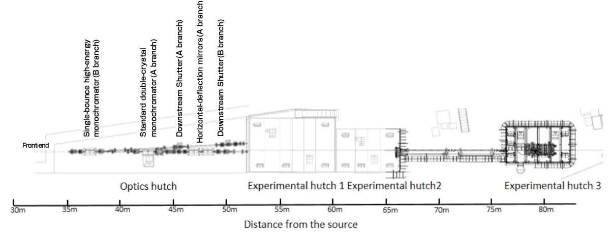 Fig.1 Schematic view of the beamline
 - X-rays beam parameters
- Branch A
Energy range 4.5 ∼ 113 keV Resolution ΔE/E 2 × 10⁻⁴ (Si (111)) Flux at sample 1012 ∼ 1013 photons/s Beam size at sample 1 (V) × 1 (H) mm2 Minimum focused beam size 100 (V)×100 (H) nm2 Higher harmonic content < 1 × 10⁻⁴
- Branch A
EXPERIMENTAL STATIONS
The beamline has three tandem experimental hutches (EH1, EH2 and EH3), which are located at 55 m, 62 m and 76 m from the source. The size of experimental hutch are 8 (L) × 5 (W) × 3.3 (H) m3, 6 (L) × 4 (W) × 3.3 (H) m3 and 6 (L) × 3.5 (W) × 3.3 (H) m3, respectively. In EH1, full-field type X-ray microspectroscopy[1] is installed (Fig. 2), In the EH2, there is a multi-purpose diffractometer and a free space for XAFS measurements using the brought-in equipment. In the EH3, scanning type X-ray microspectroscopy is installed[3] (Fig. 3). The various microspectroscopy methods supplied in BL37XU shows in Table 1.
Table 1 Microspectroscopy methods supplied in BL37XU
| Type of microscopy | Energy | Resolution | FOV | 3-dimentional | |
|---|---|---|---|---|---|
| Scanning | 4.5 ∼ 55 keV | 100 nm | µm ∼ mm | Long measurement time | |
| Full-field | projection | 4.5 ∼ 113 keV | 1 µm | mm2 | Possible |
| imaging | 6 ∼ 15 keV | 50 nm | ∼ µm | Possible | |
| Coherent diffraction | CDI | 5 ∼ 10 keV | 10 nm | µm2 | Long measurement time |
| ptychography | 5 ∼ 10 keV | 20 nm | mm2 | Long measurement time | |
To realize energy tunable X-ray nano beam, two types of KB mirror optics is adopted in X-ray microprobe system. The KB mirrors have a Rh or Pt/Rh coating, depending on the energy range used. In the EH3, the beam size was 100 (H) × 100 (V) nm2, and the photon flux was estimated to be 1010 ∼ 1011 photons/s (4.5 ∼ 15 keV), 109 ∼ 1010 photons/s (15 ∼ 37.5 keV) and 107 ∼ 108 photons/s (37.5 ∼ 55 keV)[3].
The Si 333 - Si 511 double crystal monochromator realizes higher-energy X-ray spectroscopic analysis (< 113 keV.) This enhances flexibility of high-energy XRF and enables high-energy XAFS. Figure 4 shows high-energy XRF spectra of NIST SRM610 silicate glass which is used as a reference material for various analytical methods and is doped with 61 trace elements at the 100 ppm level[4].
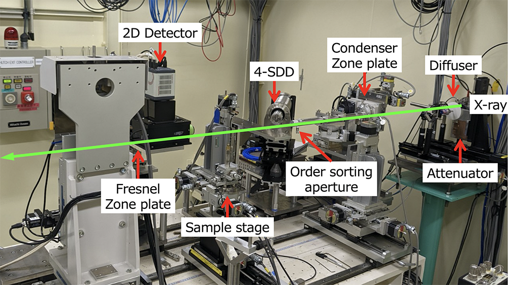
Fig.2 Full-field imaging type X-ray microspectroscopy measurement system.
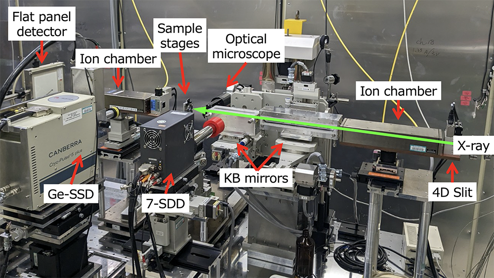
Fig.3 Scanning type X-ray microspectroscopy measurement system
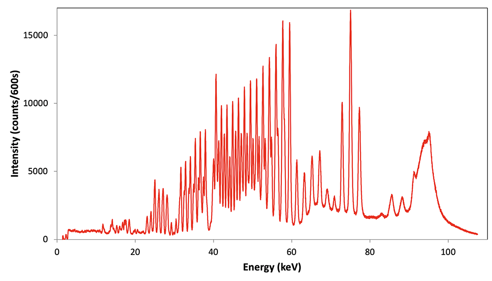
Fig.4 XRF spectrum of SRM 610 glass sample (exposure time: 600 s)
- References
- Suzuki M., Terada Y., Ohashi H., SPring-8 Research Frontiers 2011, 149-150 (2012).
- Terada Y., Tanida H., Uruga T., Takeuchi A., Suzuki Y., Goto S., AIP Conf. Proc. , 1365, 172-175 (2011).
- Ohashi H., Terada Y., et al., J. Phys: Conf. Ser. , 425, 052018 (2013).
- NItta K., Sekizawa O., SPring-8/SACLA Annual Report 2018 , 64 (2019).
- Suzuki M., Terada Y., et al., SPring-8 Riyosha Joho, 16, 201-209 (2011)[Japanese].
PUBLICATION SEARCH
* Sorry, Some parts of results are displayed using Japanese characters.
CONTACT INFORMATION
Please note that each e-mail address is followed by "@spring8.or.jp."
Oki SEKIZAWA
SPring-8 / JASRI
e-mail : sekizawa
Kiyofumi NITTA
SPring-8 / JASRI
e-mail : nittak