fig2.html
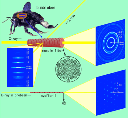
Fig. 2. Schematic diagram showing the principle of recording X-ray diffraction patterns from a muscle fiber isolated from bumblebee flight muscle (the diffraction patterns in the figures are also schematic drawings).
a: a bumblebee.
b: Diffraction recording by a conventional method (equatorial reflections. The specimen is placed perpendicular to the incident X-ray beam). Because the myofilament lattices of the myofibrils in a muscle fiber are randomly oriented, reflections originated from all lattice planes are recorded at the same time. However, the information about the orientation of the lattices is lost and the reflections having close lattice constants overlap with each other.
c: End-on diffraction recording from a muscle fiber. X-ray beams are irradiated along the fiber axis. If the beam diameter is large, the diffraction pattern consists of a set of concentric circles (powder diffraction pattern), and again the information about the orientation of the lattices is lost. This is because, in this case too, a large number of myofibrils are in the beam.
d: End-on diffraction recording from a single myofibril using X-ray microbeams. Unlike in this figure, it is not necessary to isolate a myofibril, and all that is needed is to shoot at a myofibril within the muscle fiber. Because only one single hexagonal lattice is in the beam, the reflections originating from each lattice plane appear as separate spots (This occurs only if the lattice planes in all sarcomeres are in register). If the conventional method of recording were applied to a single myofibril (Incident beams are made perpendicular to the fiber axis), it would be impossible to record all the reflections in a single exposure.