fig3.html

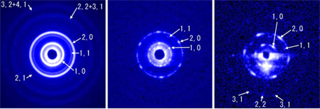
Fig. 3 End-on diffraction patterns actually recorded from the flight muscle fibers of bumblebee.
(modified from H. Iwamoto et al., Biophys. J., 83:1074-81, Fig. 2)
a: End-on diffraction pattern recorded with a 50 µm-diameter X-ray beam (about the diameter of a muscle fiber). This corresponds to the diagram in Fig. 2c. The reflections appear as concentric circles because of a large number of myofibrils in the beam.
b,c: End-on diffraction pattern recorded with a 2 µm-diameter X-ray microbeam (about the diameter of a myofibril). Reflections originating from a single hexagonal lattice appear as isolated spots. This implies that the lattices of ~1,000 sarcomeres in the ~ 3 mm beampath are perfectly in register. The dark circle at the center of the diffraction pattern is due to the beamstop to protect the detector from the direct beam.