SPring-8, the large synchrotron radiation facility

Skip to content
» JAPANESE
Personal tools
 

fig3.html

fig3-e.gif


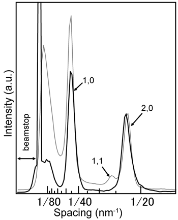
Fig. 3. Intensity profile of the diffraction pattern recorded from a single sarcomere.
Intensity profile of the diffraction pattern recorded from a single sarcomere (black solid line).
Also shown is the intensity profile obtained from an end-on diffraction pattern from a muscle fiber recorded with a 50-µm beam (gray solid line).

 
Figure 3 is reproduced with IUCr's copyright permission from "X-ray microdiffraction and conventional diffraction from frozen-hydrated biological specimens", H. Iwamoto, K. Inoue, T. Fujisawa and N. Yagi, J. Synchrotron Rad. (2005). 12, 479-483.