The World’s First Successful Internal Structure Analysis of Drug Delivery Nanoparticles (Press Release)
- Release Date
- 29 Jan, 2013
- BL40B2 (Structural Biology II)
Japan Science and Technology Agency (JST)
The University of Kitakyushu
Graduate School of Frontier Sciences, The University of Tokyo
ME Laboratory, The Jikei University School of Medicine
Japan Synchrotron Radiation Research Institute
Main points
• In spite of the high expectations placed on polymer micelles as the next-generation DDS, little has been known about the details of their internal structure.
• The first attempt ever to apply a novel variation of the anomalous small-angle X-ray scattering method on DDS particles has shed light on their accurate internal structure.
• The knowledge obtained is expected to provide basic guidelines for high-performance DDS development, leading to effective applications in gene therapy and anticancer drug delivery.
|
As part of JST Use-Inspired Fundamental Research, Prof. Kazuo Sakurai (The University of Kitakyushu) and other researchers tackled the problem of analyzing the internal structure of polymer micelle*1 particles - an agglomerated assembly of several hundreds of string-like polymeric macromolecules - which are raising expectations for use as the next-generation drug delivery system (DDS). The researchers applied the highly stable X-ray measurement system available at SPing-8*2 for the precision small-angle X-ray scattering method, and successfully revealed, for the first time in the world, how a drug is held within the particle. DDS represents a system in which a drug is delivered to the affected area selectively by encapsulating it in nanoscale carriers*4, providing advantages including: (1) a drastic reduction of side effects, (2) the potential to use a drug with untapped efficacy that has been hindered from administration because of its insolubility in water or rapid in-vivo degradation. In spite of the need to know the state of drug encapsulation for upgrading delivery performance, detailed information pertaining to internal structure has been out of reach because of the small size of DDS particles: several nanometers, or several 10-9 meters. The anomalous small-angle X-ray scattering method is a novel technique developed by a group led by Prof. Yoshiyuki Amemiya (University of Tokyo), and an effort is underway for its wider clinical application, led by researchers including Associate Prof. Masayuki Yokoyama (Jikei University School of Medicine). In this study, the method was applied for the analysis of polymer micelles. As the application of the technique to this measurement required enhanced accuracy - specifically a rigorous determination of minute variations (≤ 1%) in X-ray intensity - , Dr. Naoto Yagi (deputy director, JASRI) and other researchers employed the highly stable and intense X-ray available at SPring-8 and devised a set of special equipment to reduce noise, leading to achieving the highest level of measurement accuracy in the world. In addition, the application of the nano-interface analysis technique developed by Prof. Kazuo Sakurai et al. enabled the world’s first visualization of the internal structure of polymer micelles and how drugs are contained in it. These efforts led to the knowledge that, contrary to the conventional belief that the drug is contained inside the micelle, the drug molecules stick out of the core, forming a thin layer between the core and the shell. This knowledge is believed to carry a significant implication for the mechanism of drug release. That is, the formation of such intermediate layer while the drug is taken into the polymer micelle would enable easy drug transfer when the micelle comes into contact with a cell. The results obtained from this study are expected to help extend basic knowledge conducive to the development of higher performance DDS, leading to side effect reduction and higher performance of gene therapy and anticancer medications. The research results are slated to appear in the soon-to-be-released online version of Journal of the American Chemical Society. Publication: |
<<Figures>>
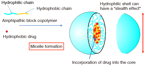
The polymer micelles are nanoparticles characterized by core-shell morphology (internal core and external shell) formed through self-association of hydrophilic and hydrophobic block copolymers. The hydrophobic internal core is capable of incorporating hydrophobic molecules, making it a candidate for DDS carrier applications for taking in drugs with poor water-solubility. The hydrophilic outer shell exerts an effect of staving off immunological rejection inside the body, resulting in a stealth effect.
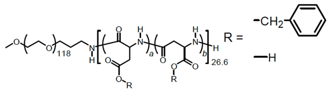
The components of the modeled polymer micelle (a block copolymer consisting of aspartate benzyl ester and polyethylene glycol). Its structure is very similar to those carriers already in the clinical stage.
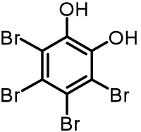
TBC (tetrabromocatechol) used as a model drug (probe molecule)
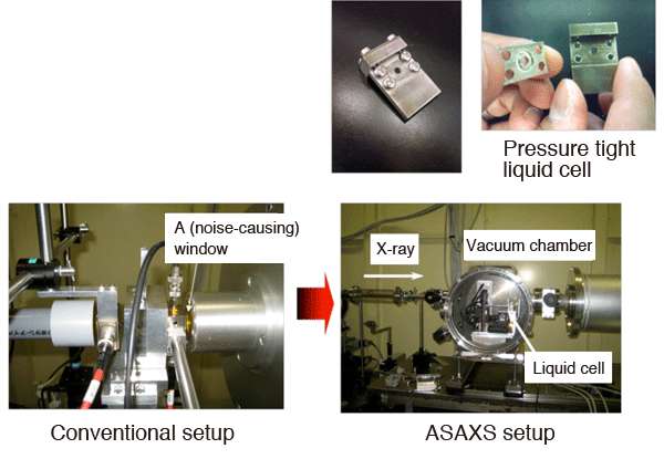
In conventional settings, samples are normally measured in air. In this study, the solution is encapsulated in a pressure tight cell (above) and placed in a vacuum chamber for measurement. This measurement configuration allows the elimination of windows, a requirement for conventional measurements in air, resulting in a drastic reduction of noise.
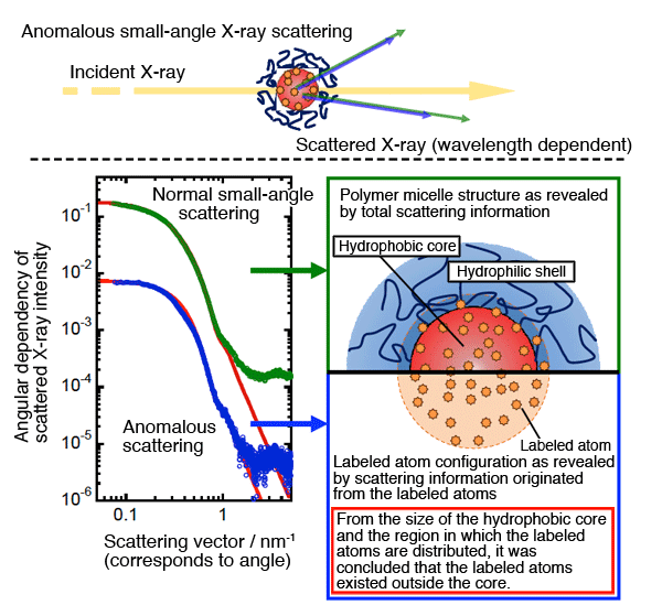
A particle irradiated by an X-ray beam scatters the beam to a varying angular range, and information concerning the angle and intensity of the scattered X-ray helps unveil the structure of the particle. If a labeled atom that absorbs an X-ray at a particular wavelength is present in the sample, it causes changes in the intensity distribution of the scattered X-ray, reflecting the state of the labeled atom. The scattering information from two sources combined - from the entire micelle and from the labeled atoms - made it possible to elucidate the structure shown in the bottom-right figure. In the figure, the labeled atoms can be seen protruding out of the hydrophobic core into the interface with the hydrophilic shell.
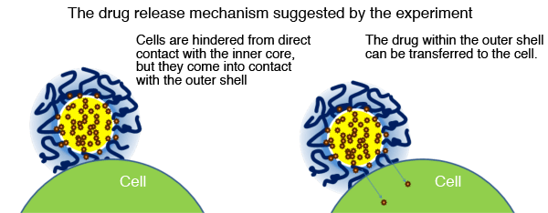
According to the conventional theory, hydrophobic drugs are considered to be encapsulated within the core of the polymer micelle. This configuration kept the mechanism of drug delivery to cells a mystery, because although shell-cell contact is possible, direct core-cell contact is unlikely. The configuration revealed by this study, in which the drug molecules are protruding into the nano-interface, means it is possible that contact between the nano-interface and a cell promotes transfer of the drug into the cell.
<<Glossary>>
*1 Polymer micelle
Nanoparticles characterized by a double-layered structure (internal core and external shell) formed through self-association of hydrophilic and hydrophobic block copolymers. Although several development examples are known, including the adriamycin (an anticancer drug) encapsulated polymer micelle, much less pharmaceutical knowledge and know-how for producing polymer micelle preparation has been accumulated than for producing tablet, granule, and powder preparations backed by massive experience. Against this backdrop, the addition of drug preparation studies such as this could drastically boost the practical use of polymer micelles.
*2 SPring-8
A RIKEN facility located in Harima Science Garden City (Hyogo prefecture) is capable of producing the world's highest intensity synchronous radiation. The management and promotion of utilization of this facility are undertaken by JASRI. The name “SPring-8” comes from “Super Photon ring-8GeV.” An electron flying at nearly the speed of light, if deflected from its original trajectory through the effect exerted by a magnet, emits an electromagnetic wave in a direction tangential to its trajectory, which is called radiation light (or synchrotron radiation). At present, there are three “3rd Generation” large scale synchronous radiation facilities in the world: SPring-8 (Japan), APS (USA) and ESRF (France). The acceleration energy available at SPring-8 (8 billion electron volts) enables the provision of an extremely wide spectrum of radiation light: from far infrared to visible, vacuum ultraviolet, and soft X-ray up to hard X-ray. SPring-8 provides a theater for collaborative works involving researchers inside and outside Japan, and the research conducted at this facility cover such diverse areas as material science, geoscience, life science, environmental science, and various applications in industrial sectors.
*3 Anomalous small-angle X-ray scattering (ASAXS)
The small-angle X-ray scattering (SAXS) method is known for its capacity to provide structural information in the nanoscale domain, and thus is widely used as a powerful analysis method for nanotechnology. As compared to SAXS, which uses a single incident wavelength, ASAXS is characterized by its use of multiple wavelengths, which enables the obtaining of information pertinent to a particular element.
*4 Carrier
The material that is used in a drug discovery system to incorporate drugs and acts as a vehicle to deliver them to the target site. A variety of carriers are being studied for their purpose-oriented suitability: type of drugs, targeted regions, and required efficacy period. These carriers provide great leeway for the design of various functions, allowing for a broad spectrum of applications. Polymer micelles constitute a class of promising carrier materials.
|
For more information, please contact:
Associate Prof. Masayuki Yokoyama (The Jikei University) |


- Current article
- The World’s First Successful Internal Structure Analysis of Drug Delivery Nanoparticles (Press Release)