Successful Nanoscale Observation of Living Cells Using X-ray Laser (Press Release)
- Release Date
- 07 Jan, 2014
- SACLA
Hokkaido University
RIKEN SPring-8 Center
Japan Synchrotron Radiation Research Institute (JASRI)
Tokyo University of Pharmacy and Life Sciences
Institute of Environmental Microbiology, Kyowa-kako Co., Ltd.
Key points
• High-contrast imaging of internal nanostructures of living cells using X-ray free-electron laser (XFEL)
• Successful visualization of natural-state living cells, immediately before damage by radiation, using X-rays with femtosecond pulse duration
• Offering insights for understanding phenomena in living cells and clarifying nanostructures of natural-state biological molecules
|
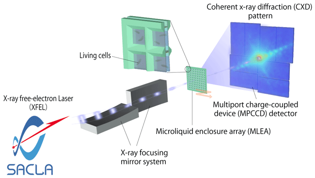
Scientists from Hokkaido University, RIKEN SPring-8 Center, JASRI, Tokyo University of Pharmacy and Life Sciences, and the Institute of Environmental Microbiology, Kyowa-kako Co., Ltd., observed living cells at the nanoscale using the SPring-8 Angstrom Compact Free-Electron Laser (SACLA)*1, an XFEL facility. The research group was led by Yoshinori Nishino (professor) and Takashi Kimura (assistant professor) from the Research Institute for Electronic Science, Hokkaido University; Yoshitaka Bessho (team leader, currently visiting scientist) from RIKEN SPring-8 Center; and Yasumasa Joti (team leader) from JASRI. Conventionally, it has been impossible to observe living cells at nanometer (one-billionth of a meter) resolution by electron microscopy or X-ray microscopy. This is because cells will die when exposed to the electron beams or X-rays used for observation. The research group succeeded in visualizing the appearance of living cells, immediately before damage by radiation, using XFEL with an extremely short pulse duration of 10 femtoseconds*2 or shorter. An advanced method called coherent X-ray diffraction (CDX)*3 was used for observation, and high-contrast images of intracellular nanostructures were obtained. The results of this research revealed that XFEL has great potential for observing biological samples close to their natural state. In the future, XFEL will be increasingly applied to cell biology. By further improving the resolution of the technique, the scientists will also pave the way for key medical application of XFEL, such as the clarification of nanostructures of natural-state biological molecules. This research was partially supported by the Ministry of Education, Culture, Sports, Science and Technology through the XFEL Priority Strategy Program; by the Japan Science and Technology Agency through the Core Research for Evolutionary Science and Technology (CREST); and by the Japan Society for the Promotion of Science through a Grant-in-Aid for Scientific Research. The achievements of this research were published online in the British scientific journal Nature Communications on 7 January 2014. Publication: |
<<Figures>>
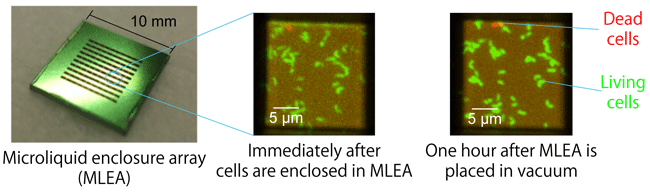
enclosure array (MLEA) can maintain living cells in the natural state
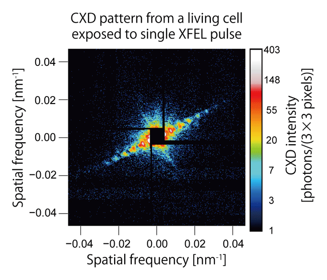
cell exposed to a single XFEL pulse
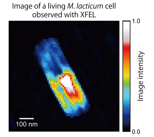
obtained by numerically analyzing CXD pattern measured using XFEL
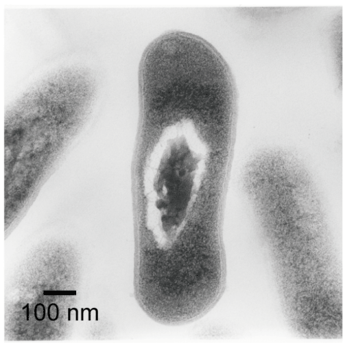
The cells were subjected to complicated treatment: they were fixed with resin, sliced into sections less than 100 nm thick, and stained with heavy-metal salts, such as uranyl acetate and lead citrate.
<<Glossary>>
*1 SACLA, an XFEL facility
Japan’s first XFEL facility constructed jointly by RIKEN and JASRI. As one of the five national critical technologies in the Basic Program for Science and Technology in Japan, the facility was constructed and developed in a five-year project starting from fiscal 2006. It was completed in March 2011 and named SACLA after the initial letters of SPring-8 Angstrom Compact Free Electron LAser. The first successful generationof an X-ray laser was in June 2011. Shared operation started in March 2012. Since then, SACLA has been used in various experiments. Although the facility is smaller than those in other countries, SACLA can produce lasers with the world’s shortest wavelength of 0.1 nm or shorter.
*2 femtosecond
A femtosecond is one-quadrillionth of a second. In one femtosecond, light (speed, ≈300,000 km/s) travels only 0.3 μm; that is, a femtosecond is an extremely short duration.
*3 Coherent X-ray diffraction (CXD)
Coherent light refers to light waves that are in phase and is a characteristic of laser light. When a specimen is irradiated with coherent X-rays, the X-rays scatter; this is CXD. CXD patterns are sensitive to even a slight difference in the structure of specimens. Numerical analysis of CXD patterns can produce images of specimens.
|
For more information, please contact: |

- Previous Article
- Analysis of Ancient Artworks Using SPring-8 (Press Release)
- Current article
- Low Core-Mantle Boundary Temperature Inferred from the Solidus of Pyrolite (Press Release)