Synchrony of beating between implanted iPS cells and the host heart confirmed at a molecular level -a significant advance for cardiac regenerative medicine using iPS cells- (Press Release)
- Release Date
- 23 Jan, 2015
- BL40XU (High Flux)
Osaka University
National Cerebral and Cardiovascular Research Center
Japan Synchrotron Radiation Research Institute
Important points
•Confirmation of synchrony between implanted iPS cells and a host heart
•Important evidence for development of the next generation therapy of cardiac disease
•Combination of two leading-edge techniques: synchrotron X-ray diffraction developed by NCVC and SPring-8, and cardiac regeneration with iPS cells.
|
Cardiac muscle regeneration with iPS cells is regarded as the next-generation cardiac therapy. To realize this, evaluation of effects of the therapy is necessary. In this research, Professor Hideki Sawa, Associate Professor Shigeru Miyagawa, Assistant Professor Satsuki Fukushima of Graduate School of Medicine, Osaka University, Dr. Mikiyasu Shirai of National Cerebral and Cardiovascular Research Center, and Dr. Naoto Yagi of Japan Synchrotron Research Institute, in collaboration with Dr. James Pearson of Monash University and Australian Synchrotron, demonstrated in an animal experiment with a highly advanced X-ray nano-diffraction technique that the contractile proteins in cells that derived from transplanted iPS cells work in synchrony with cardiac muscle cells of the host heart. Acknowledgement: Original report: |
1. Background of research
Cardiac muscle regeneration therapy with iPS cells is expected to be the next generation therapy of heart failure. In fact, it has been reported in animal experiments conducted in several countries that function of an infarcted heart can be improved by transplantation of self-beating cells that are induced to differentiate to cardiac muscle cells from iPS cells. However, it has not been proven that the transplanted cells contribute to improvement of cardiac function by contracting in synchrony with the host heart under an electric connection, which is the most important mechanism in cardiac regeneration.
2. Methods and findings
The research group demonstrated that contractile proteins in cardiac muscle cells that were transplanted to an infarcted heart operate in synchrony with the host heart, which was shown by recording X-ray diffraction from a beating rat heart using an intense X-ray beam of SPring-8. This method enables evaluation of activities of cardiac contractile proteins, which are the source of contractile force of the heart, at various points in a ventricle. The method has been already announced by National Cerebral and Cardiovascular Institute and Japan Synchrotron Radiation Research Institute on the 5th of January 2013.
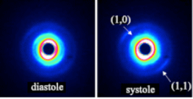
In the present study, infarction was induced by suturing left coronary artery of a rat heart. Then, a sheet of cultured iPS cells that were directed to cardiac muscle cells was transplanted onto the heart. When the cells merged with the host heart, X-ray diffraction peaks from contractile proteins in the cells were recorded using intense X-rays of SPring-8 (fig.1).
The intensity change of these diffraction peaks was in synchrony with the contraction of the cardiac muscle, showing that the contractile proteins are working in synchrony with the heart and producing force. This result shows that the cells derived from iPS cells are working as a part of the heart, and it is expected that cardiac function is improved by regeneration of cardiac muscle cells.
3. Significance of this study
It is very important to clarify the mechanism of effects of the therapy to realize cardiac muscle regeneration therapy with iPS cells. In this work, it was possible to capture the contraction of iPS cells-derived cardiac muscle cells that were transplanted to a heart using the cutting-edge synchrotron diffraction technique.
This work provided important evidence for the development of next-generation therapy for cardiac diseases by combining two leading-edge techniques, iPS cells and SPring-8, that originated in Japan. It is anticipated that science will be advanced by a combination of techniques in different scientific fields, as demonstrated in this work.
<<Figures>>
|
For more information, please contact:
Graduate School of Medicine, National Cerebral and Cardiovascular Research Center, Japan Synchrotron Radiation Research Institute, |
- Previous Article
- Discovering New Autoimmune Disease Mechanism -Success in x-ray single molecule observations of Autoimmune System- (Press Release)
- Current article
- Synchrony of beating between implanted iPS cells and the host heart confirmed at a molecular level -a significant advance for cardiac regenerative medicine using iPS cells- (Press Release)