High-precision tracking of ultrafast molecular oscillations -Creating molecular movie with atomic-level spatiotemporal resolution-
- Release Date
- 09 Aug, 2019
- SACLA
August 9, 2019
Japan Synchrotron Radiation Research Institute
RIKEN
High Energy Accelerator Research Organization
|
A joint research team led by Tetsuo Katayama (research scientist) of the XFEL Utilization Division, Japan Synchrotron Radiation Research Institute (JASRI); Makina Yabashi (group director) of the Beamline Research and Development Group, RIKEN SPring-8 Center; Professor Shin-ichi Adachi of the Institute of Materials Structure Science, High Energy Accelerator Research Organization; Christian Bressler (group leader) of the European XFEL (Germany); Christopher Milne (group leader) of the Paul Scherrer Institute (Switzerland); Professor György Vankô of the Hungarian Academy of Sciences (Hungary); and Professor Thomas Penfold of Newcastle University (UK) has succeeded in tracking nuclear wavepacket oscillations*1 in a photoexcited metal complex with an atomic-level high spatiotemporal resolution, using the X-ray free-electron laser (XFEL)*2 at SACLA*3. They created a “molecular movie” for observing and understanding the “motion of molecules during a photoreaction”. Their achievement is an important step in clarifying the photoreaction mechanisms. The Cu(I)-phenanthroline complex, which is a promising photosensitizer*4, is known for its structural change from a regular tetrahedron to a flattened structure upon light absorption. The joint research team studied how nuclear wavepacket oscillations are associated with this structural change by time-resolved X-ray absorption spectroscopy*5 using an XFEL. The XFEL, which realizes both a pulse duration of a hundred-trillionth of a second and a wavelength of ten-billionth of a meter, can produce a molecular movie by clearly capturing the structure of the photoexcited Cu(I)-phenanthroline complex without blurring. As a result, they observed three types of nuclear wavepacket oscillations during the relaxation process. One is the breathing motion associated with the stretching and shrinking of the Cu-N bond lengths. The remaining two are bending vibrations in which the bond angles between Cu and N atoms change. They found that the lifetimes of these nuclear wavepacket oscillations are different, and that the two types of bending vibrations are strongly associated with the structural flattening of the Cu(I)-phenanthroline complex. Their research achievements have been published online in the international scientific journal Nature Communications.
|
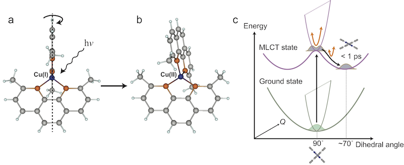
Fig. 1 Structural flattening of Cu(I)-phenanthroline complex.
(a) Regular tetrahedral structure before light irradiation. (b) Flattened structure after light irradiation. When the Cu(I)-phenanthroline complex absorbs light, the dihedral angle between the two phenanthroline ligands is reduced from 90° to ~70°. (c) Potential energy surface landscape of the Cu(I)-phenanthroline complex absorbing light and changing structure while oscillating.
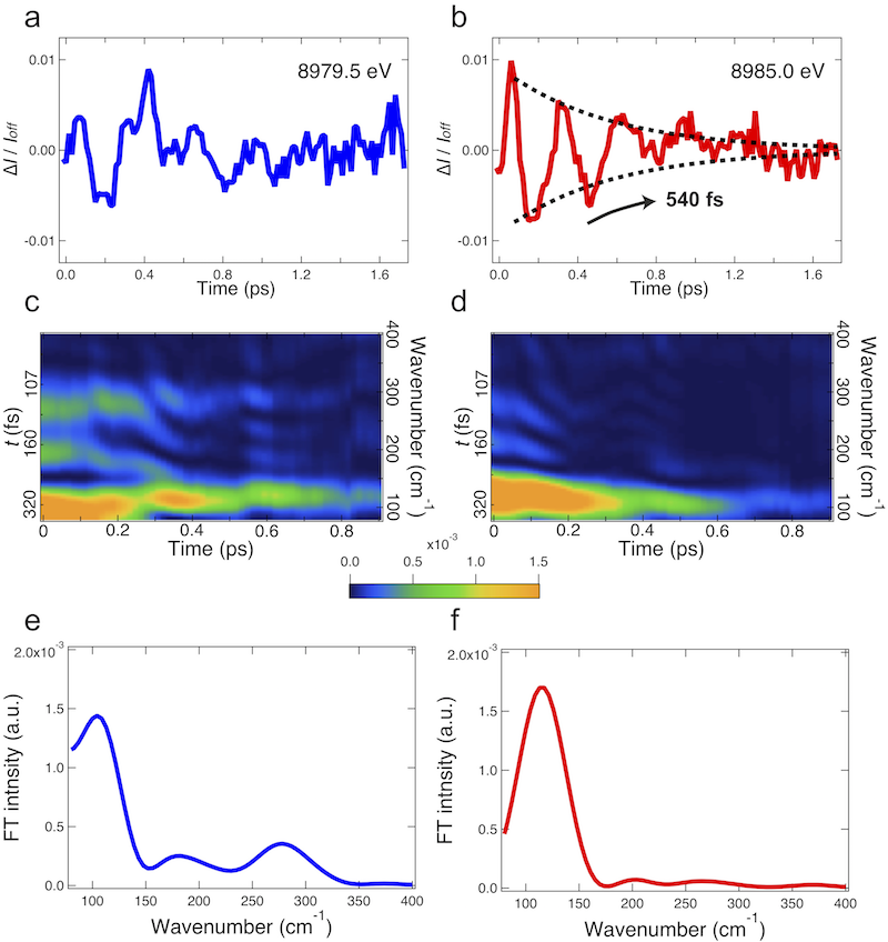
Fig.2 Nuclear wavepacket oscillations in photoexcited Cu(I)-phenanthroline complex.
(a,b) Oscillations observed at incident X-ray energies of 8979.5 and 8985.0 eV, respectively. Time 0.0 indicates the time when a visible laser pulse is irradiated onto the complex molecules. One picosecond (ps) is one-trillionth of a second. (c,d) Mapping of Fourier analysis. Colors indicate the intensity of nuclear wavepacket oscillations at each time・during the relaxation process. (e,f) Integrated Fourier transforms within the time range from 0 to 0.4 ps. While three vibrational modes are observed at 8979.5 eV (c,e), only a single mode is observed at 8985.0 eV (d,f). The peak at ~100 cm-1 corresponds to a breathing vibration, whereas the oscillations at ~180 cm-1 and ~280 cm-1 correspond to bending vibrations. In (c), the bending vibrations disappear at approximately 0.2 ps, which is earlier than the disappearance of the breathing vibration. These results support that the two bending vibrations make a large contribution to the structural flattening of the Cu(I)-phenanthroline complex.
Glossaries
※1.Nuclear wavepacket oscillations
When multiple waves with different wavelengths are superposed, the amplitude increases where the peaks and troughs coincide but decreases where the peaks and troughs annihilate each other. As a result, temporal or spatial localization with a large amplitude occurs (a wavepacket). The nuclear wavepacket is generated by the superposition of vibrational wave functions of a substance.
※2.X-ray free-electron laser (XFEL)
A pulse laser in the X-ray wavelength region realized by the recent advancement of accelerator technology. Unlike conventional lasers using semiconductors and gases as the oscillation medium, an XFEL uses electron beams that travel at a high speed in vacuum and theoretically has no restrictions on the wavelength used. An XFEL can produce pulses a billion times brighter than those produced by conventional synchrotron radiation sources such as SPring-8 on a femtosecond (one-quadrillionth of a second) time scale. Taking advantage of the extraordinarily high brilliance, an XFEL is used for the structural analysis of proteins with atomic resolution using nanometer-scale crystals, as well as for the clarification of nonlinear optical phenomena in the X-ray region.
※3.SACLA
Japan’s first XFEL constructed jointly by RIKEN and JASRI. SACLA is one of the five national core technologies in Japan’s Third Science and Technology Basic Plan. The five-year project for the construction and preparation of SACLA was launched in FY2006 and was completed in March 2011. The name SACLA is short for SPring-8 Angstrom Compact free electron LAser. In June 2011, the first oscillation of the X-ray laser was achieved. Since March 2012, SACLA has been open to public users. SACLA is capable of generating lasers with the world’s shortest wavelength of ≤0.1 nm.
※4.Photosensitizer
A substance that absorbs light and transfers its energy to other substances in order to assist the progress of a chemical reaction.
※5.Time-resolved X-ray absorption spectroscopy
An X-ray is absorbed by an atom at energies near and above the core-level binding energies of that atom. X-ray absorption spectroscopy is an experimental technique for measuring the absorbance of samples by varying the incident X-ray wavelength and can provide information on the local structure and chemical state in the vicinity of atoms of interest. Time-resolved X-ray absorption spectroscopy is a technique for studying the time evolution of a fast phenomenon by tuning the timing of short-duration X-ray (from an XFEL) and visible laser pulses.
Contact: |
 spring8.or.jp
- Previous Article
- Fight Against Global Warming – NTU Multi-Nation Research Published on《Science》(Press Release)
- Current article
- High-precision tracking of ultrafast molecular oscillations -Creating molecular movie with atomic-level spatiotemporal resolution-