Development of High-Resolution In Vivo-CT System for Small Animals Adopting High-Brilliance Synchrotron Radiation as Light Source - First ever three-Dimensional Dynamic Observation of Peripheral Airway and Coronary Artery of Living Animals(Press Release)
- Release Date
- 12 Aug, 2008
- BL20B2 (Medical and Imaging I)
Key research achievements
- Visualization of peripheral airway and coronary artery (125 μm in diameter) of living mice by synchrotron radiation at SPring-8
- Realization of not only three-dimensional stationary but also three-dimensional dynamic observation (four-dimensional observation)
-
Substantial contribution not only to pharmacology and physiology but also to living-matter simulation
RIKEN (Ryoji Noyori, President) has developed a high-resolution in vivo-CT system that enables the three-dimensional dynamic analysis of the peripheral airway and coronary artery of small living animals (mice and rats) using the Medical and Imaging I Beamline BL20B2 of SPring-8. This is the achievement of the members of the following research group: Toshihiro Sera and Hideo Yokota, the scientists of the Living Matter Simulation Research Team (Ryutaro Himeno, team leader) of the RIKEN Advanced Science Institute (Kohei Tamao, Director); Naoto Yagi, the deputy director of the Research & Utilization Division, Japan Synchrotron Radiation Research Institute (JASRI; Akira Kira, Director General); and Hiroyuki Tachibana, the instructor of the Department of Medical Engineering, Faculty of Health Science and Technology, Kawasaki University of Medical Welfare.
Gene therapy and regenerative medicine have attracted attention, and much effort is directed towards developing new drugs. With the aim of applying these drugs to clinical practice, animal experiments before clinical trials to determine the efficacy and ensure the safety of new drugs are very important. Small animals are often used in animal experiments before clinical trials because they are suitable not only for testing the efficacy and safety of drugs but also for manipulating specific genes. Images of the small living animals are a must to follow the time course of the effects of drugs on organs with periodic large beating, such as the heart and lungs. In this case, it is important to realize high-resolution three-dimensional dynamic observation to catch the minute changes caused by the drugs with a wide range of view.
Recently, various three-dimensional dynamic observation systems for small animals, using visualization devices such as X-ray CT, MRI and PET, have been proposed. But, for small living animals, the time available for capturing images is very severely limited because their heartbeat and breathing rhythms are approximately six to ten times faster than those of humans. Accordingly, it was difficult to obtain detailed contrast using conventional systems. In conventional systems, only large tissues such as the airways and bronchial tubes of the lungs and the ventricles and atrium of the heart can be visualized. This research team has therefore focused on the beamline at SPring-8 having a brilliance approximately one hundred million times higher than that of a conventional X-ray generator and has developed a high-resolution in vivo-CT system with a high-intensity synchrotron radiation as a light source. In this system, the time period for capturing an image is synchronized with the heartbeat and breathing to reduce motion artifacts due to the heartbeat and breathing. As a result, the researchers successfully realized the three-dimensional dynamic observation of the peripheral airway, coronary artery and aortic valve, which are approximately 125 μm in diameter, of living mice, that cannot be captured by conventional systems.
We hope that the drug response test using the developed system and the simulations of living body and treatment using the results of the test will be applied to a wide range of fields, including physiology and pharmacology, and the consideration of treatment strategy.
This research achievement was published in the British scientific journal, Physics in Medicine and Biology (the August 21, 2008 issue).
Publication:
"Development of high-resolution 4D in vivo-CT for visualization of cardiac and respiratory deformations of small animals"
Toshihiro Sera, Hideo Yokota, Kazuhiro Fujisaki, Kazuaki Fukasaku, Hiroyuki Tachibana, Kentaro Uesugi, Naoto Yagi and Ryutaro Himeno
Physics in Medicine and Biology 53, 4285-4301 (2008), published online 24 July 2008
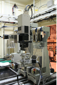 High-resolution in vivo-CT system
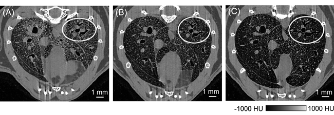 Dynamic observation of the peripheral airway of a living mouse by the high-resolution in vivo-CT system:
(A) at a pressure of 0 cmH2O (B) at a pressure of 5 cmH2O (C) at a pressure of 15 cmH2O. The white circle indicates the same bronchial tube.
|
For more information, please contact: |

- Current article
- Development of High-Resolution In Vivo-CT System for Small Animals Adopting High-Brilliance Synchrotron Radiation as Light Source - First ever three-Dimensional Dynamic Observation of Peripheral Airway and Coronary Artery of Living Animals(Press Release)