The Development of an Image Analysis Method for High-Resolution Observation of the Beating Heart: Looking into the Dilation/Contraction Mechanism of the Coronary Artery and Cardiac Muscle in Mouse Models of Cardiovascular Disease (Press Release)
- Release Date
- 05 Jan, 2013
- BL28B2 (White Beam X-ray Diffraction)
- BL40XU (High Flux)
‹New technology that helps elucidate the molecular mechanism of cardiovascular diseases and accelerates the development of therapeutic methods›
National Cerebral and Cardiovascular Center (NCVC)
Japan Synchrotron Radiation Research Institute (JASRI)
Monash University
University of Otago
|
The researchers at NCVC (director: Nobuo Hashimoto, Suita city, Osaka pref.) *, JASRI (director: Tetsuhisa Shirakawa, Sayo town, Hyogo pref.) **, Monash University***, and University of Otago**** have teamed up and conducted a high-intensity X-ray-assisted study on observation and elucidation of the detailed behavior of beating hearts in mice and rats commonly used for modeling cardiovascular diseases. The main objectives of the study include observation of the dilation and contraction mechanism of the tiny coronary artery, and the elucidation of motor functions of proteins that drive myocardial contraction, whereby the high-intensity synchrotron radiation X-ray available at SPring-8 played an integral role. The team has developed technologies - many of them are world firsts - along the way and elucidated molecular mechanisms of cardiovascular disease. The US specialty journal Circulation Research highly acknowledged these achievements and published a review article in its online publication on the 4th of January. The technologies developed in this study concern image analysis of small-animal hearts that are rapidly beating: 1) synchrotron radiation assisted high resolution microangiography capable of observing whole view of coronary responses, from the main artery to arterioles (down to approximately 30-50 μm in diameter), 2) a synchrotron radiation X-ray diffraction method that enables multiple pinpoint assessments of contractile protein movements - the source of cardiac contraction and relaxation - in different regions of the heart. These technologies are expected to pave the way for elucidating molecular mechanisms, involving genes and proteins, that cause cardiovascular diseases, and for accelerating therapeutic method development. * Dr. Mikiyasu Shirai (director: Department of Cardiac Physiology), et al. Reference: |
<<Figures>>
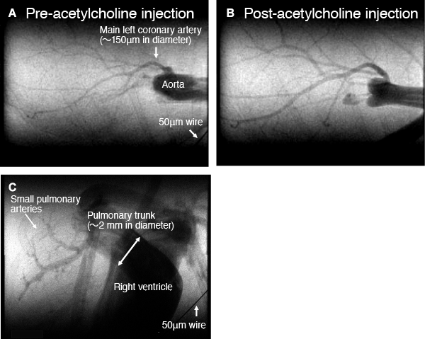
(heart rate: up to 500 beats/min): the coronary arteries (A, B) and the right ventricle and pulmonary arteries (C)
The small size of the mouse heart permits coronary imaging of the whole heart, from the main artery to the arterioles with a diameter of 30-50μm (A). Intravenous injection of acetylcholine (endothelium-dependent vasodilator) dilated the coronary arteries in the wide area (B). Fast image acquisition of the mouse lung enables visualization from the right ventricle to the pulmonary trunk and even to small pulmonary arteries and estimation of pulmonary blood flow transit time (C).
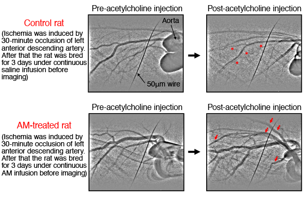
post-ischemic-reperfusion small coronary arteries of rats.
Coronary microangiography was performed in the in situ rat heart 3 days after ischemia-reperfusion injury. Single-frame images of iodine contrast during baseline condiotion (left two figures) and during acetylcholine infusion (right two figures) in a vehicle-treated control (upper two figures) and AM-treated (0.05 μg/kg per minute continuous infusion for 3 days by osmotic pump) rat (lower two figures). AM-treated rat heart showed vasodilation (indicated by red arrows) and vessel recruitment during acetylcholine infusion, whereas control rat showed abnormal vasoconstriction (indicated by “*”) and loss of vessels. Ischemia was induced by 30-minute occlusion of the left anterior descending artery under anesthesia 3 days before imaging.
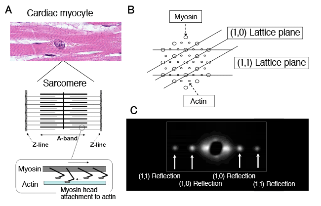
A: Magnified view of the cardiac myocyte striation showing the sarcomere structure. Its A-band has interdigitating filaments consisting of actin and myosin. During contraction, the myosin head moves and binds to actin, generating contractile force.
B: Cross-section of the A-band in striation. Thin actin and thick myosin filaments form a hexagonal lattice arrangement. In this lattice arrangement, the (1, 0) lattice plane consists solely of myosin, and the (1, 1) lattice plane consists of actin and myosin.
C: An X-ray diffraction image of cardiac muscle. The inner pair represents reflections from the (1, 0) lattice planes, and the outer pair from the (1, 1) lattice planes, called (1, 0) and (1, 1) reflection, respectively. During myocardial contraction, the myosin head moves to actin, causing mass transfer from the (1, 0) to (1, 1) lattice plane. The mass transfer increases (1, 1) reflection and decreases (1, 0) reflection, resulting in a reduced intensity ratio of these reflections, i.e. I1,0/I1,1. During myocardial relaxation, the reverse process takes place, resulting in increased I1,0/I1,1 ratio.
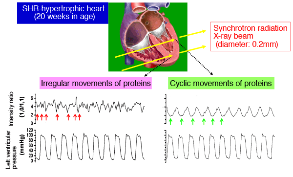
left ventricular pressure, for 10-pulse period, in a spontaneously hypertensive rat (SHR).
In a normal myocardial region, the myosin head regularly repeats binding to, and dissociation from actin. The cyclic change in intensity ratio reflects this reversible process. The signal obtained in the anterior wall region showed normal cyclic changes in sync with heart rate, but that in the posterior wall region showed irregularities (indicated by red arrows). This finding shows that the functional failures of contractile proteins in myocardial contraction and relaxation do not develop uniformly throughout the heart, but nonuniformly from region to region. The synchrotron radiation X-ray diffraction method is the only noninvasive method permitting multiple pinpoint (dimensions 0.2x0.2 mm) assessments of myocardial function at the molecular level in different regions of the heart.
|
For more information, please contact: |
- Current article
- The Development of an Image Analysis Method for High-Resolution Observation of the Beating Heart: Looking into the Dilation/Contraction Mechanism of the Coronary Artery and Cardiac Muscle in Mouse Models of Cardiovascular Disease (Press Release)