Development of Innovative X-ray Microscopy with High Spatial Resolution and Sensitivity (Press Release)
- Release Date
- 04 Mar, 2013
- BL29XU (RIKEN Coherent X-ray Optics)
Osaka University
RIKEN
Key research findings
• Visualization of a slight phase shift of approximately 1/320 of the X-ray wavelength at a spatial resolution of approximately 10 nm
• Expected application to the observation of biological samples with a high spatial resolution using SPring-8 facilities and the SPring-8 Angstrom Compact Free-Electron Laser (SACLA)
|
A research group led by Yukio Takahashi (associate professor) of the Graduate School of Engineering, Osaka University, and Tetsuya Ishikawa (chief scientist) of RIKEN SPring-8 Center succeeded in developing an X-ray microscopy method with high sensitivity and spatial resolution. An object with even little light absorption can be visualized by observing the shift in the phase*1 generated when X-rays pass through the object. This technique is called phase-contrast imaging. The research group developed a new phase-contrast imaging method called X-ray ptychography along an optical illumination system that combines an X-ray converging mirror and a spatial filter*3 using the RIKEN physics beamline I (BL29XUL) at SPring-8.*2 The research group succeeded in visualizing a small phase shift of approximately 1/320 of an angstrom-order X-ray wavelength with a high resolution of approximately 10 nm by X-ray ptychography. This microscopy is particularly effective for observing biological soft tissues composed of low-mass elements with little light absorption. The technique is expected to be applied to various bioimaging methods using SPring-8 facilities. In addition, a similar optical system has been adopted in a coherent X-ray diffraction imaging experiment using the X-ray free-electron laser SACLA.*4 Imaging with ultimately high sensitivity and high spatial resolution is expected to be realized using X-ray lasers with a high photon density such as SACLA. The achievement of this study was published online in Applied Physics Letters on 4 March 2013. Publication: |
<<Figures>>
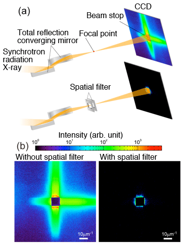
(b) intensity distribution taken by charge-coupled device (CCD)
Synchrotron radiation X-rays with an energy of 8 keV are focused on a 100-nm-diameter spot using a total reflection converging mirror. Extra X-rays scattered from the total reflection converging mirror can be effectively eliminated by placing a rectangular opening slit with an aperture size of approximately 100 μm near the focal point as a spatial filter.
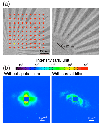
Red dots indicate the positions of X-ray irradiation.
(b) Diffraction pattern observed when the position indicated
by a yellow dot is irradiated with X-rays
A custom-made test chart manufactured by NTT Advanced Technology Corporation was used as the sample. A thin tantalum film of approximately 12 nm thickness was micromachined to obtain a minimum structure of 17 nm size. The results of comparing the intensity distribution taken by the CCD with and without a spatial filter upon the irradiation of X-rays indicate that the intensity distribution of X-rays scattered from the sample can be measured with a high signal-to-noise ratio by adopting the spatial filter.
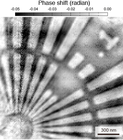
by focused X-ray ptychography with spatial filter
A phase shift of approximately 0.02 rad is visualized. The spatial resolution is estimated to be approximately 10 nm because the minimum structure with a size of 17 nm is resolved.
<<Glossary>>
*1 Phase
A phase refers to a position within a waveform that periodically changes, and a wavefront is the locus of points having the same phase. A wave can be represented by the amplitude of the wavefront and the phase. The amplitude of a light wave that passes through an object attenuates and a phase shift occurs. Therefore, the attenuation of the wave amplitude is related to the absorption contrast, whereas the phase shift is related to the phase contrast.
*2 SPring-8
SPring-8 is a shared facility that delivers the most brilliant synchrotron radiation currently available in the world, and is located at Harima Science Park City, Hyogo Prefecture. The name “SPring-8” is derived from “Super Photon ring-8 GeV”. Synchrotron radiation is a type of light produced when charged particles are forced to travel in a curved path by a magnetic field. SPring-8 can produce X-rays with a high coherence because of the small size of the circulating electron groups and their high stability.
*3 Spatial filter
A spatial filter is a minute hole inserted in the spectral plane of an optical system to emphasize a particular spatial frequency or suppress high-frequency noise.
*4 X-ray free-electron laser, SACLA
SACLA is a next-generation generator of X-rays that are completely coherent. In Japan, the generator was built in the campus of SPring-8 by RIKEN in cooperation with Japan Synchrotron Radiation Research Institute (JASRI). The generator, called SACLA, was completed in March 2011. The first successful oscillation of the X-ray laser was in June 2011, shared operation started in March 2012, and since then SACLA has been used in various experiments.
|
For more information, please contact: |

- Previous Article
- Magnesium binding and sarcolipin regulation of calcium pump as revealed by X-ray crystallography (Press Release)
- Current article
- Development of Innovative X-ray Microscopy with High Spatial Resolution and Sensitivity (Press Release)