Approach to Understanding Thermostable Toxin of Vibrio parahaemolyticus That Causes Food Poisoning
Large-scale food-poisoning epidemic 60 years ago
In October 1950, the most widespread food-poisoning epidemic in the postwar period occurred in the southern area of Osaka Prefecture affecting 292 people and killing 20 of them. The patients began complaining of stomachache and had diarrhea and vomiting about an hour after they ate dried whitebait (Shirasu-boshi), and the late Tsunesaburo Fujino [professor emeritus of the Research Institute for Microbial Diseases (RIMD), Osaka University] identified Vibrio parahaemolyticus contaminating dried whitebait as the causal pathogen for the food poisoning. Since then, the students of Professor Fujino have taken over research on V. parahaemolyticus, which was the first food-poisoning-causing bacterium discovered in Japan.
Dr. Itaru Yanagihara, a student of Professor Fujino, has continued his research on V. parahaemolyticus since he moved from RIMD, Osaka University, to Osaka Medical Center and Research Institute for Maternal and Child Health. He succeeded in clarifying the three-dimensional structure of thermostable direct hemolysin (TDH), a toxin protein produced by V. parahaemolyticus, at the atomic level in May 2010. This was achieved through joint research with Takeshi Honda, a professor emeritus of RIMD, Osaka University, and Hiroshi Hashimoto, an assistant professor of Yokohama City University.
Their achievement is attracting attention because it will lead to the clarification of the mechanism of food poisoning.
V. parahaemolyticus - a cunning bacterium
Professor Fujino described V. parahaemolyticus as “plump and stubby.” It is a rod-shaped bacterium of approximately 2 μm (a micrometer is one-millionth of a meter) in length with one thick flagellum and thin flagella (as shown in the image on the cover). Using these flagella for locomotion, the bacterium swims freely in the sea and adheres to fish and shellfish. At this time, the number of bacterial cells is not large enough to cause food poisoning. However, the bacterium divides once every 10 minutes at optimum temperature and salt concentration. At this rate of proliferation, a bacterial cell divides into 64 bacterial cells in an hour and after 6 hours there will be 68.7 billion bacterial cells. At this high cell concentration, the bacterium causes food poisoning.
Once the bacterium gains entry into the human body and adheres to the intestinal tract, it casts a secretion apparatus similar to a ship's anchor in order to transfer proteins into the body. It eliminates other types of bacteria from the intestine and colonizes the intestinal tract. Moreover, it secretes a toxin that destroys the cardiac muscle cells of the host. The toxin is called TDH (Photograph 1) because it is thermostable and, at the time of discovery, was found to form a pore on the membrane of red blood cells, which ruptures these cells.
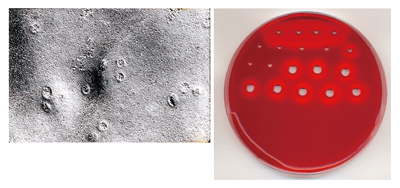
Photograph 1 Pores formed by TDH on cell membrane (left) and view of hemolysis (right)
On an agar plate containing blood, hemolysis occurs where TDH is dropped, forming a clear zone.
(Source of photograph on left: Toshio Miwatani and Takeshi Honda, Visual Materials for Education of Bacteriology (second edition) published by Japanese Society for Bacteriology)
Toxicity not eliminated by heating
“We sometimes say we can eat old food if it is sufficiently heated. But it's not the case with V. parahaemolyticus,” says Dr. Yanagihara. Generally, proteins that are denatured by heating do not return to their original state. For example, a boiled egg does not return to its raw state. That is why we think heated food is safe. However, in the case of V. parahaemolyticus, although the bacterium is killed by heating, the toxicity of TDH, which is once lost, may be regained depending on the subsequent temperature changes. This is called the Arrhenius effect named after the discoverer of this phenomenon. The late Toshio Miwatani, a professor emeritus of RIMD, Osaka University, found for the first time in 1972 that TDH produced by V. parahaemolyticus showed the Arrhenius effect, but the detailed mechanism underlying this effect in this bacterium was not clarified.
The toxicity of TDH is determined by its structure
Dr. Yanagihara began to focus on the Arrhenius effect of TDH when he found that TDH combined with β globulin (a general type of protein contained in milk). When β globulin was fixed on the membrane of an experimental device, TDH combined with β globulin. However, when TDH was fixed on the membrane, β globulin did not combine with TDH. Dr. Yanagihara assumed that this was because the three-dimensional structure of TDH was destroyed when TDH was fixed on the membrane. Namely, the structure determines the characteristics of TDH.
Then, Dr. Yanagihara evaluated the toxicity of TDH following its structural changes depending on temperature using various measurement devices (Fig. 1). In his experiments, TDH was found to have a fibrous structure (TDHi)*1 and an unfolded structure (TDHu) in addition to its toxic structure (TDHn). Dr. Yanagihara and his colleagues were very much delighted about this finding and concluded, “The Arrhenius effect of TDH can be explained by its structure.”
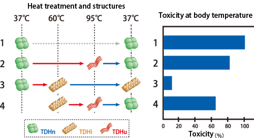
Fig. 1 Arrhenius effect of TDH produced by V. parahaemolyticus
The right figure shows the toxicity remaining after heat treatment, assuming the toxicity before treatment (TDHn; 1) as 100%. The left figure shows the structural changes in TDH. When TDH is heated at 60°C (3), it changes its structure to the fibrous structure (TDHi) and its toxicity is not regained even when it is cooled. When TDH is heated at a temperature higher than 60°C, it changes its structure to an unfolded structure (TDHu) and loses its toxicity; however, when it is cooled to body temperature, it is refolded (TDHn) and the toxicity is regained (2 and 4).
Is it really a tetramer?
Through experiments on the structure of TDH, they began to think that toxic TDH forms a tetramer.*2 Because the toxin had been considered a dimer, an atomic-level analysis was absolutely necessary to prove that toxic TDH is a tetramer. Therefore, in order to directly observe the structure of TDH, X-ray crystallography of crystallized TDH was performed with the cooperation of Dr. Hahsimoto of Yokohama City University (Fig. 2). The analysis was performed using the BL41XU and BL40B2 beamlines of SPring-8 and a beamline of the High Energy Accelerator Research Organization (KEK). The structural analysis of TDH was attempted about 10 years ago but it was unsuccessful. Dr. Hashimoto says, “It is not easy to produce crystals of high quality with a suitable size for analysis. However, we succeeded in doing this to some extent in previous experiments. The radiation experiment technology at SPring-8 is upgraded every year. Things that used to be invisible have become visible.” The key to the success in their experiment was the improvement of measurement technology.
With the clarification of the three-dimensional structure of TDH, the research project made substantial progress. A pore with a diameter of approximately 2 nm (a nanometer is one-billionth of a meter) and a depth of approximately 5 nm was observed at the center of the tetramer of TDH. Water molecules were observed to rapidly pass through this pore in a molecular simulation using a supercomputer, and it is considered that this pore leads to the formation of a pore on the cell membrane of the host.
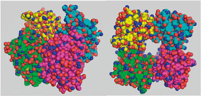
Fig. 2 Crystal structure of TDH
Structure of tetramer (left): Assembly of four color-coded chains.
Overhead view (right): A pore penetrates through the center of the tetramer. Each ball indicates an atom.
Final stretch of research on TDH
“What should be further clarified is how TDH approaches the cell membrane of the host and forms a pore to exert its toxicity,” says Dr. Yanagihara whose research project will now enter the final stretch. He has already determined a model of TDH in his mind; however, he thinks that it will remain only a model unless it is verified. “In order to verify the model, we need a radiation strong enough to enable the structural observation of a substance that is not crystallized.” He looks forward to the development of SPring-8 and the introduction of the X-ray free-electron laser (XFEL), which will soon be operational.
As a result of 60 years research on V. parahaemolyticus, people understand the importance of food administration, and the examination method to detect V. parahaemolyticus has been established, leading to the significant decrease in the number of patients affected by V. parahaemolyticus. Still, the successors of Professor Fujino, the discoverer of this bacterium, are carrying out research with the aim of “eradicating infection by V. parahaemolyticus,” a hope of Professor Fujino.
Glossary
*1 Fibrous structure
A fibrous structure is a row of molecules in a rodlike, long, thin shape.
*2 Tetramer
Proteins are polymers consisting of chains of numerous amino acids. When multiple chains assemble by binding and work as one, the resulting compound is called a multimeric protein. When a compound consists of one chain, two chains, or four chains, it is called a monomer, dimer, or tetramer, respectively. Each chain is called a subunit.
Column: Surrounded by good colleagues
 |
| Dr. Yanagihara with one of his colleague Dr. Hashimoto (on the left) |
“To approach a problem that has not been solved through decades of research in RIMD, we need the power of science and engineering,” says Dr. Yanagihara. Indeed, researchers from various fields were involved in this research. All of them were young researchers in their mid-30s in neighboring laboratories when Dr. Yanagihara was in RIMD, and Dr. Hashimoto was also among them. Arguing with them over one thing or another and making free use of the laboratory equipment of the engineering department at night when nobody else was using it are pleasant memories for Dr. Yanagihara.
After moving to the Osaka Medical Center and Research Institute for Maternal and Child Health, Dr. Yanagihara was able to continue his research on V. parahaemolyticus while working as a pediatrician. He is grateful to the people around him who have been understanding and supportive of his research activity.
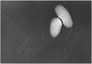 |
Image on cover Electron micrograph of V. parahaemolyticus, which causes food poisoning
Bacterial cells and thick flagella are seen in this image. |
Interview and original text by Akiko Ikeda (Sci-Tech Communications Incorporated)
This article was written following an interview with Itaru Yanagihara, the head of the Department of Developmental Medicine, Research Institute, Osaka Medical Center for Maternal and Child Health, and Hiroshi Hashimoto, an assistant professor of the Graduate School of Nanobioscience, Yokohama City University.