Local structure analysis of rewritable optical media during phase change

問い合わせ番号
SOL-0000001053
ビームライン
BL01B1(XAFS I)
学術利用キーワード
| A. 試料 | 無機材料 |
|---|---|
| B. 試料詳細 | 金属・合金, 半導体, 結晶性固体, 非晶質、ガラス, 薄膜(無機) |
| C. 手法 | 吸収、及びその二次過程 |
| D. 手法の詳細 | XAFS, EXAFS, XANES |
| E. 付加的測定条件 | 偏光(直線), 室温 |
| F. エネルギー領域 | X線(4~40 keV) |
| G. 目的・欲しい情報 | 結合状態, 局所構造, 構造変化, 機能構造相関, 機能発現, 相転移 |
産業利用キーワード
| 階層1 | 記憶装置 |
|---|---|
| 階層2 | CD-R、DVD |
| 階層3 | |
| 階層4 | 液体・非晶質構造, 原子間距離, 結晶構造, 局所構造, 電子状態 |
| 階層5 | XAFS |
分類
A80.12 半導体・電子材料, A80.30 無機材料, M40.10 XAFS
利用事例本文
In this solution, EXAFS method was applied to Ge2Sb2Te5 thin film (20 nm thickness), the material of rewritable optical media, to analyze local structural change during phase change. EXAFS spectra of the sample in crystalline and amorphous phase were measured at Ge, Sb and Te K-edge. The EXAFS method is a powerful technique to study local structure (distance, coordination number, species of neighbor atoms) of selected elements both in crystalline states and in non-crystalline states. Figure 1 shows radial structure function of Te atom after Fourier transform of EXAFS spectra. Simultaneous analysis of EXAFS data set at 3 edges revealed that the fast rewritable mechanism is due to the change in position of Ge atom in rigid building blocks.
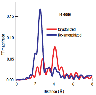
Fig. 1 Radial structure function for Te atom in crystallized and amorphized samples.
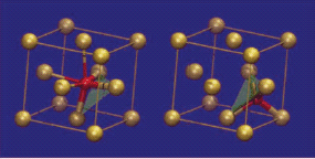
Fig. 2 Fragments of the local structure of GST around Ge atoms in the crystalline (left)
and amorphous (right) states (red: Ga).
[ A. V. Kolobov, P. Fons, A. I. Frenkel, A. L. Ankudinov, J. Tominaga, T. Uruga, Nature Materials 3, 703-708 (2004), Fig. 1(c), 5,
©2004 Nature Publishing Group ]
画像ファイルの出典
原著論文/解説記事
誌名
Nature Materials, 3, 703 (2004)
図番号
1(c), 5
測定手法
XAFS spectra of thin film samples having a high density of core-hole atoms are obtained by measuring Auger electron yield from excited atoms as a function of x-ray energy. A conversion electron yield (CEY) detector is used for this measurement. The CEY XAFS mode is successfully applied for the 0.1 nm thick film samples. Acquisition time per spectrum is 1-2 hr.
画像ファイルの出典
私信等、その他
詳細
講習会プレゼン資料
測定準備に必要なおおよその時間
4 時間
測定装置
| 装置名 | 目的 | 性能 |
|---|---|---|
| XAFS Measurement System | Measurement of XAFS spectra | 3.8-113 keV |
| CEY Detector | Measurement of XAFS spectra of dense thin film | thickness > 0.5 nm |
参考文献
| 文献名 |
|---|
| A. Kolobov et al., Nature Materials, 3, 703 (2004). |
関連する手法
アンケート
SPring-8だからできた測定。他の施設では不可能もしくは難しい
本ビームラインの主力装置を使っている
測定の難易度
中程度
データ解析の難易度
熟練が必要
図に示した全てのデータを取るのにかかったシフト数
1シフト以下