BL04B1 X-ray radiography
Inquiry number
INS-0000000351
A high-resolution CCD camera is attached to the large-volume press so that we can see the macroscopic change of the sample shape under high pressure and high temperature using an X-ray radiography technique (Fig. 1). The use of the X-ray radiography technique does not only make it easy to adjust the X-ray beam to the desired position in the sample, but has also led to the development of new techniques to observe dynamic processes under high-pressure, such as sample deformation, melting. As is shown in fig. 1, the incident white X-ray from the bending magnet irradiates the sample cell through the anvil gap, and an image of the sample is projected on the fluorescence screen. This image is then magnified and detected by a high-resolution CCD camera. Real-time images of the sample are captured and recorded in a computer. Recently, a new X-ray radiography system combining with a high-magnification lens and a high-speed CCD camera was installed, and each image data can be captured at intervals of 1/125 second with the resolution of less than 4 μm/pixels (Fig. 2). The high-speed X-ray radiography system has been applied to the viscosity experiments of the melts under pressure using the “falling sphere” technique.
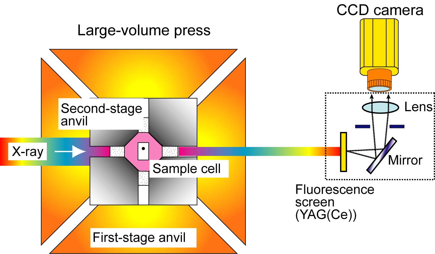
Fig. 1. A schematic drawing of the X-ray radiography system on the large-volume press.
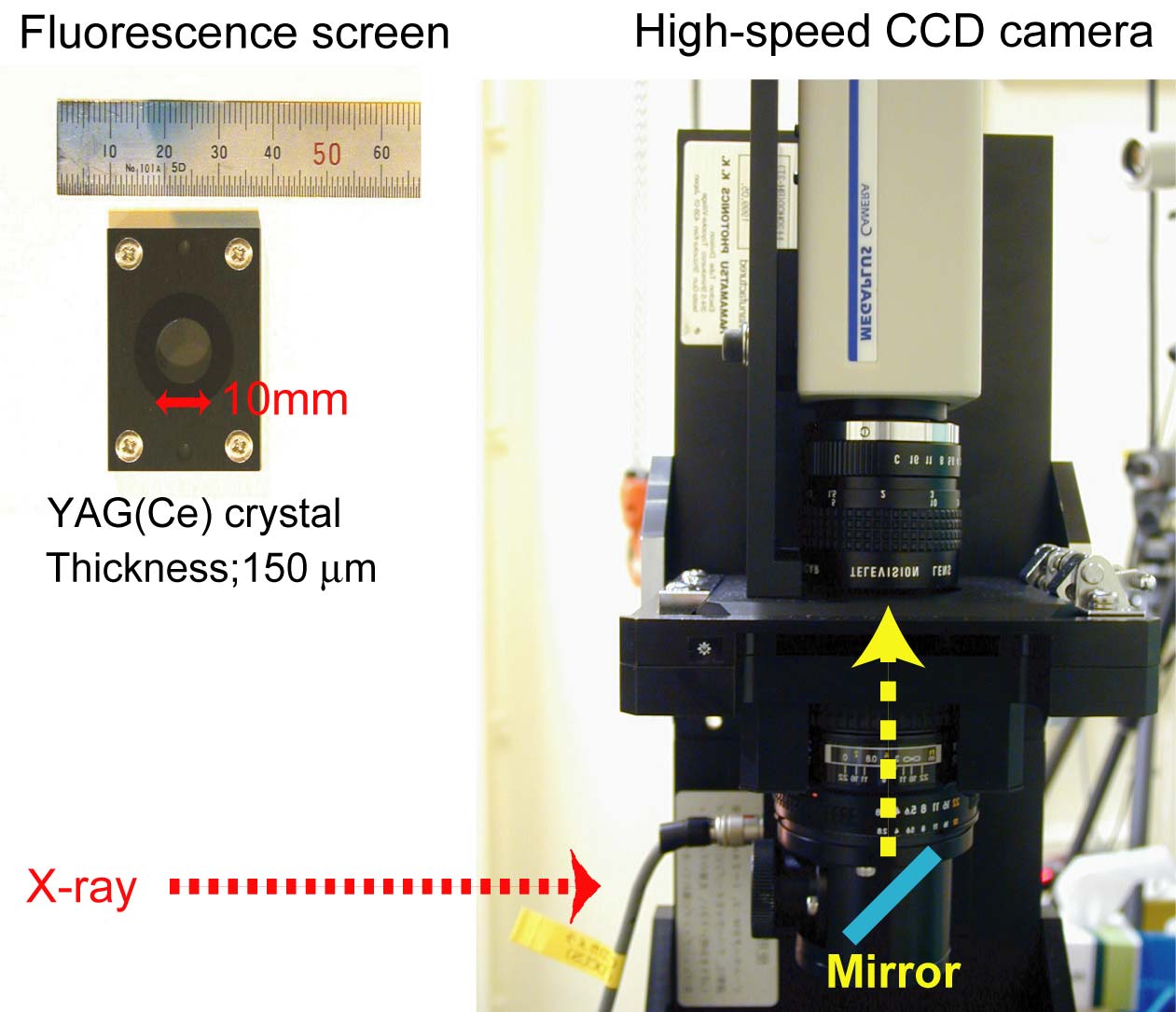
Fig. 2. New X-ray radiography system combining with a
high-magnification lens and a high-speed CCD camera.