Determination of the second critical endpoint in silicate–H2O system

問い合わせ番号
SOL-0000001195
ビームライン
BL04B1(高温高圧)
学術利用キーワード
| A. 試料 | 無機材料 |
|---|---|
| B. 試料詳細 | 絶縁体・セラミックス, 液体・融体, 溶液 |
| C. 手法 | 透過X線計測, 吸収、及びその二次過程 |
| D. 手法の詳細 | |
| E. 付加的測定条件 | 二次元画像計測, 高圧(大型プレス), 応力付加、変形, 高温(>>500度) |
| F. エネルギー領域 | X線(>40 keV) |
| G. 目的・欲しい情報 | 構造変化, 相転移 |
産業利用キーワード
| 階層1 | 環境, 工業材料, その他 |
|---|---|
| 階層2 | |
| 階層3 | |
| 階層4 | 内部構造, 形態 |
| 階層5 | イメージング |
分類
A80.30 無機材料, A80.90 その他
利用事例本文
H2O plays an important role in magmatism and chemical evolution in Earths interior. Two types of hydrous mobile phase, aqueous fluid and hydrous silicate melt, could exist in Earths crust and mantle. In general, both the solubility of water in silicate melt and the solubility of silicate materials in aqueous fluid increase with increasing pressure. This could suggest that, above a certain pressure and temperature, silicate melt and aqueous fluid become indistinguishable from each other. This critical condition is called the second critical endpoint. The use of X-ray radiography technique using a large volume press, SPEED-1500 has enabled the direct observations of coexisting aqueous fluid and silicate melt. As is shown in figure, the spherical bubble were observed in the SrAl2Si2O8-H2O system from 3 GPa to 4 GPa, whereas no bubbles were observed during heating up to about 1500℃ at 4.3 GPa. These results first indicate that aqueous fluid and melt can coexist at pressures up to 4 GPa, and the second critical endpoint exists at pressure between 4and 4.3 GPa in the SrAl2Si2O8-H2O system.
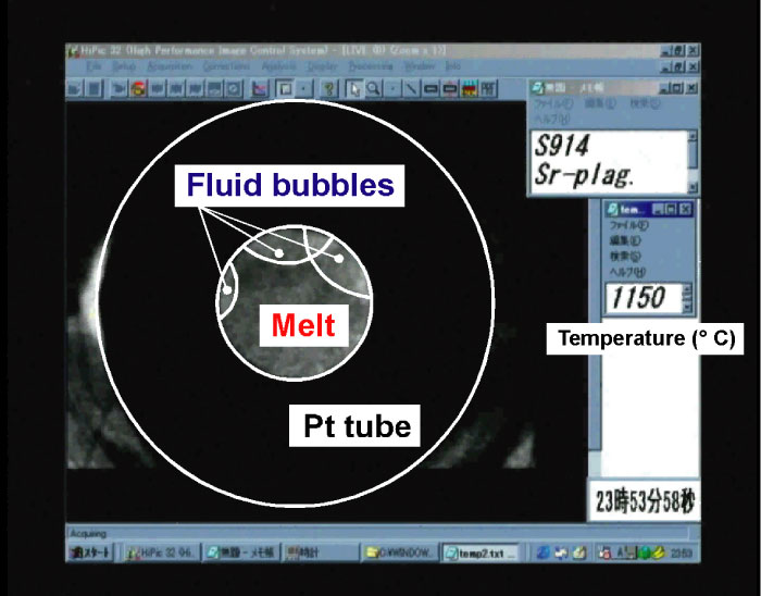
Fig. X-ray radiographic image of the SrAl2Si2O8-H2O system at 3 GPa and 1150℃.
[ K. Mibe, M. Kanzai, T. Kawamoto, K. Matsukage, Y.-W. Fei and S. Ono, Geochimica et Cosmochimica Acta 68, 5189-5195 (2004), Fig. 5,
©2004 Elsevier, Ltd. ]
画像ファイルの出典
所内報
誌名
Research Frontiers (2003)
ページ
88
測定手法
A high-resolution CCD camera is attached to the large-volume press so that we can see the macroscopic change of the sample shape under high pressure and high temperature using an X-ray radiography technique (Fig. 1). The use of the X-ray radiography technique does not only make it easy to adjust the X-ray beam to the desired position in the sample, but has also led to the development of new techniques to observe dynamic processes under high-pressure, such as sample deformation, melting. As is shown in fig. 1, the incident white X-ray from the bending magnet irradiates the sample cell through the anvil gap, and an image of the sample is projected on the fluorescence screen. This image is then magnified and detected by a high-resolution CCD camera. Real-time images of the sample are captured and recorded in a computer. Recently, a new X-ray radiography system combining with a high-magnification lens and a high-speed CCD camera was installed, and each image data can be captured at intervals of 1/125 second with the resolution of less than 4 μm/pixels (Fig. 2). The high-speed X-ray radiography system has been applied to the viscosity experiments of the melts under pressure using the “falling sphere” technique.
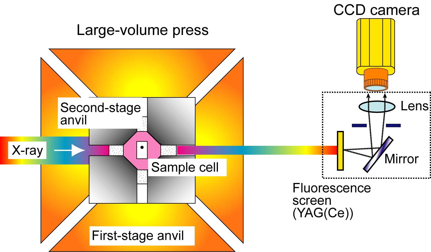
Fig. 1. A schematic drawing of the X-ray radiography system on the large-volume press.
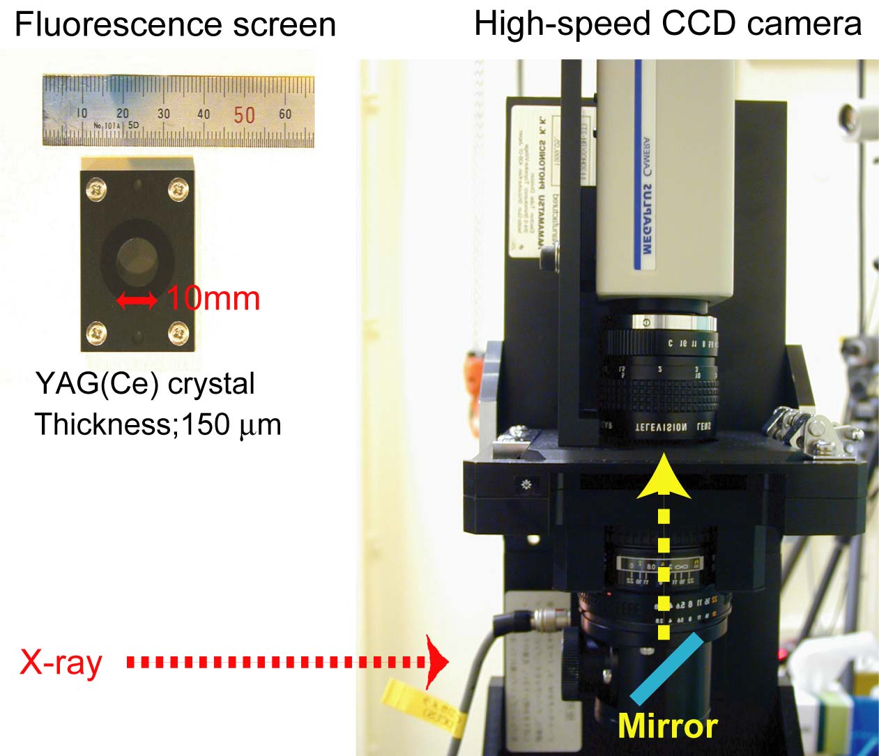
Fig. 2. New X-ray radiography system combining with a high-magnification lens and a high-speed CCD camera.
画像ファイルの出典
BL評価レポート
ページ
16
測定準備に必要なおおよその時間
1 日
測定装置
| 装置名 | 目的 | 性能 |
|---|---|---|
| SPEED-1500 | High pressure and high temperature experiment | 2500 K, 30 GPa |
参考文献
| 文献名 |
|---|
| Geochimica et Cocmochimica Acta, 68, 5189(2004) |
関連する手法
アンケート
SPring-8だからできた測定。他の施設では不可能もしくは難しい
本ビームラインの主力装置を使っている
測定の難易度
中程度
データ解析の難易度
中程度
図に示した全てのデータを取るのにかかったシフト数
2~3シフト