BL10XU OUTLINE
Inquiry number
INS-0000000352
ABSTRACT
Beamline BL10XU is dedicated to high-pressure research using diamond anvil cells (DACs). We can perform high-pressure powder x-ray diffraction under the conditions of high-pressure and a wide range of temperature from cryogenic region to thousands of kelvins. A major feature of this beamline is high-brilliance and high-energy X-rays produced from synchrotron radiation source of the in-vacuum type undulator. The monochromatic and focused X-ray beam (6∼61 keV) irradiates samples enclosed in DACs which enable the generation of high-pressures over 300 GPa. The XRD Debye-Scherrer rings from the sample are collected on an image plate (IP) area detector, an X-ray flat panel detector (FPD), or high speed CdTe hybrid pixel array detector. In experimental stations, cryostats and a double-sided laser heating system have been prepared to provide the extreme temperature conditions. By combining high pressure generation using DACs with the use of high-energy, high-brilliance X-rays of SPring-8, users cover a wide range of scientific research fields such as high-pressure condensed matter physics, materials sciences, mineral physics, and Earth and planetary sciences, and specialize in in-situ measurements of material properties under the extreme conditions: determination of crystal structure, pressure-induced structural phase transitions, and equation of state, and the analysis of phase diagram.
X-RAY SOURCE AND OPTICS
A high-intensity, high-energy, and quasi-monochromatic X-ray beam is supplied from an in-vacuum type undulator. The undulator beam is monochromatized by a SPring-8 standard type double crystal monochromator (DCM) with liquid-nitrogen cooling, resulting in intense X-rays (>1013 photons/s) with a sufficient energy resolution (ΔE/E ∼ 10-4). The DCM is equipped with an Si (111) crystal and an Si (220) crystal, which covers X-rays energy to 37 keV and 61 keV, respectively.
The stacked compound X-ray refractive lenses (CRL), which are made from glassy carbon or aluminum, are installed at a downstream position of 4.3 m from the DCM. The CRL device provides high spatial and angular resolutions (about 10 arcseconds) suitable for X-ray diffraction experiments at sample position. Using CRL in optics hutch, the X -ray spot size is 0.05 mm (V) × 0.15 mm (H) at sample position (FWHM). About 7 times intensity gain is obtained by CRL when X-rays energy is tuned at 30 keV. By a multi-stage focusing method in combination with a refractive lens installed in the experimental hutch, we can finally provide a micro-focused beam with 0.8 µm (V) × 0.9 µm (H) at the sample position.
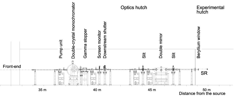
Fig. 1. Schematic view of BL10XU optics hutch
- MAJOR FEATURE OF BL10XU
Available x-ray energy 6 - 61 keV Energy resolution ΔE/E ∼10-4 X-ray beam size φ 0.001 - 1.0 mm Flux ∼1.0 × 10¹³ photons/sec (30 keV), ∼1.0 × 10¹² photons/sec (61 keV)
EXPERIMENTAL STATIONS
The equipment for high-pressure x-ray diffraction experiments are installed in a tandem-type experimental hutch in BL10XU. High-pressure study with DACs is conducted utilizing x-ray energies from 6 to 61 keV.
EXPERIMENTAL HUTCH1
A low vibration He-closed-cycle GM cryostat has been installed to perform high pressure and low temperature XRD measurements with oscillations. A helium gas driven membrane system in the cryostat can be used to compress samples in DAC remotely. An on-line Raman spectroscopy system with a 532 nm diode laser is equipped for pressure measurements at cryogenic temperature. The system consists of a microscope unit with a long working distance objective lens and a silver-coated glassy carbon mirror (Fig. 2). The glassy carbon mirror is employed as laser delivery optics and is transparent to high energy X-rays. X-ray diffraction images are collected on an image plate area detector (IP, Rigaku. Co., R-AXIS IV++; 300×300 mm2; pixel size 0.1 mm × 0.1 mm) or a Perkin Elmer digital X-ray flat panel detector (FPD, XRD0822 CP23; 1024×1024 pixels; pixel size 0.2 mm × 0.2 mm). The camera distance between a sample and the detector is tunable between 220 and 540 mm and between 200 and 450 mm, depending on a measuring range of diffraction angle and an angular resolution.

Fig. 2. Integration system of X-ray diffractometer and Raman scattering for high pressure and low temperature experiments in experimental hutch 1
EXPERIMENTAL HUTCH2
High-pressure X-ray diffraction experiments can be carried out using the laser-heating system and micro X-ray beam. Incident X-ray optics, a diffractometer for DACs, an X-ray detector stage, and laser heating optics have been installed, which are mounted on independent optical tables (Fig. 3). The optical tables on which the incident x-ray optics and laser heating optics are mounted have two translation stages, allowing precise alignment of these optics to the X-ray beam position. The sample stage system, which consists of high-precision and high-load X-Y-Z translation and rotation stages, is designed for mounting the various DACs (e.g., a cube type DAC for ultra-high- pressure experiments and a symmetric piston-cylinder type DAC for laser heating).
The XRD Debye rings are collected on the Varex Imaging digital X-ray flat panel detector (FPD, XRD1611 CP3; 4096×4096 pixels; pixel size 0.1 mm × 0.1 mm). The sample-to-detector distance for FPD can be shifted between 270 and 470 mm. High speed CdTe hybrid pixel array detector (LAMBDA CdTe 750k, X-Spectrum GmbH; pixel size 55 µm × 55 µm, detector pixels 512×1528) is also available. On the detector stage, a microscope unit is equipped to observe samples in DACs and to align the sample to X-ray beam. The microscope unit can also be used as Raman probe head for on-line pressure measurements and Raman spectroscopy measurement.
A double-sided laser heating system with near-infrared fiber lasers is installed for high-pressure and high-temperature X-ray diffraction experiments to study mineral physics of deep Earth materials and to synthesize new high-pressure materials. Fig. 4 shows the laser heating system for DACs installed in the BL10XU. The Ag-coated glassy carbon mirror is employed to deliver the laser to the sample in the DAC and is transparent over 90% for high-energy x-ray above 30 keV. For the purpose of generating high temperatures (above 5000 K) under high pressure, two TRUMPF fiber lasers (100 W) have been installed since 2024. The laser spot size on the sample can be selected ranging from 15 to 100 µm.
In experimental hutch 2, the low-vibration helium-closed-cycle GM cryostat has been installed for low-temperature X-ray diffraction experiments using a micro-focused beam. A helium gas driven membrane system in the cryostat can be used for remotely applying pressure on a sample in the DAC.
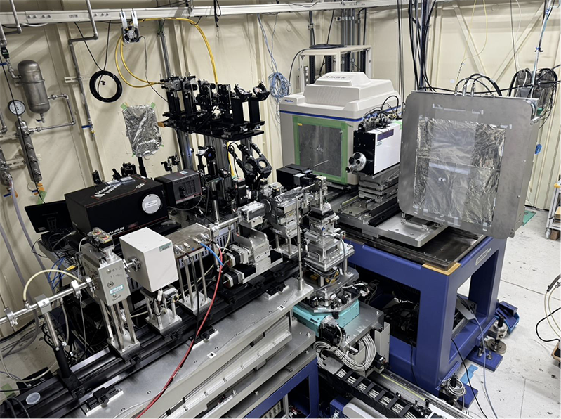
Fig. 3. High-precision diffractometer system for high-pressure x-ray
diffraction experiments equipped in the experimental hutch 2.
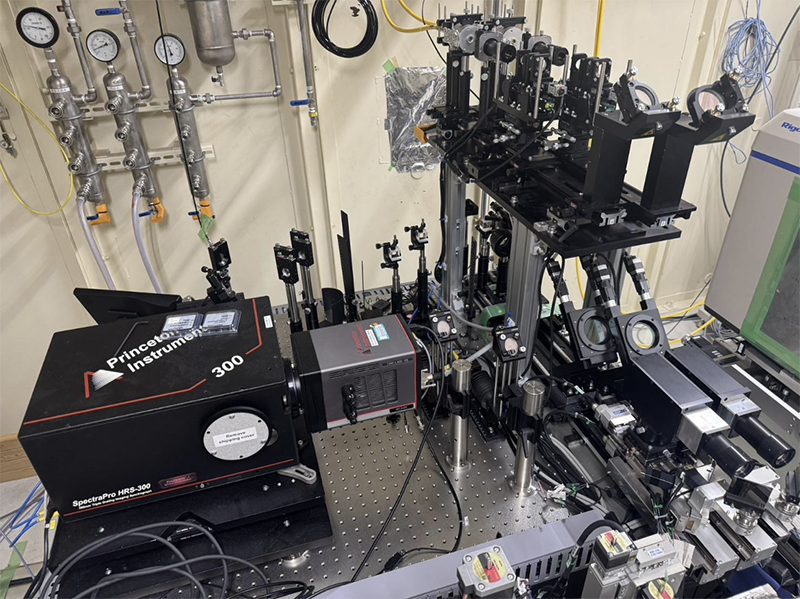
Fig. 4. The optics of double-sided laser heating and temperature
measurements in the experimental hutch 2
DATA EVALUATION SOFTWARE
The IP detector, R-AXIS IV++, and the FPD are operated using an X-ray diffraction data acquisition software designed by Rigaku Co. and a programming software in LabVIEW, respectively. Stages for sample and detector and optics for Raman spectroscopy system and laser heating system can be controlled from LabVIEW software. For Raman data collection and processing, Teledyne Princeton Instruments LightField software is available.
EQUIPMENT HIGHLIGHTS
- Image plate area detector (IP)
- X-ray flat panel detector (FPD)
- CdTe hybrid pixel array detector
- Micro Raman spectroscopy system for pressure measurements (on-line): 532 nm diode lasers (hutch 1, 2)
- Electrical resistance measurement system
- Cryostat and thermostat (∼10 K)
- Double-sided laser heating system (1500 ∼ 6000 K)
- Diamond anvil cell (Bring your own)
- Sample preparation room in experimental hall
- Electric discharge-drilling machine (EDM)
- Ruby fluorescence system for pressure measurement (off-line): 488 nm diode laser
- Micro Raman spectroscopy system for pressure measurements (off-line): 785 nm diode laser
- Micro Raman spectroscopy system (off-line): 532 nm diode laser
PUBLICATION SEARCH
* Sorry, Some parts of results are displayed using Japanese characters.
CONTACT INFORMATION
Please note that each e-mail address is followed by "@spring8.or.jp."
Naohisa HIRAO
Japan Synchrotron Radiation Research Institute (JASRI)
1-1-1 Kouto, Sayo-cho, Sayo-gun, Hyogo 679-5198
Phone : +81-50-3495-0798
e-mail : hirao
Hirokazu KADOBAYASHI
Japan Synchrotron Radiation Research Institute (JASRI)
1-1-1 Kouto, Sayo-cho, Sayo-gun, Hyogo 679-5198
Phone : +81-50-3496-9089
e-mail : kadobayashi
Sho KAKIZAWA
Japan Synchrotron Radiation Research Institute (JASRI)
1-1-1 Kouto, Sayo-cho, Sayo-gun, Hyogo 679-5198
e-mail : kakizawa