BL20B2 Image detectors
Inquiry number
INS-0000000315
Image detectors
THigh-spatial-resolution 2-dimensional image detectors are prepared for radiographic imaging. The detectors are a fluorescent-screen lens-coupling system (Fig.8). X-rays passing through the object are transformed into a visible image by the fluorescent screen. Images on the screen are read by a cooled CCD camera with a high numerical aperture lens.
The CCDs consist of respectively (Table). The equivalent pixel sizes projected onto the screen area are 5.83 µm when C4880-10-14A is combined with coupling lenses (beam monitor AA40), which have the magnification factor of 2. The equivalent pixel size is 5.87 µm when C4742-95HR is combined with a lens (beam monitor AA60), which has the magnification factor of 1.
The high-spatial resolution CCD image detectors take images of biological specimens by using techniques of micro-tomography and refraction-contrast imaging. Digitized images with 14 or 12 bits resolution are captured and stored into a personal computer. These detectors are also used for novel imaging techniques using various kinds of optical elements.
A real-time digital micro-imaging system is under development for micro-angiography so that small blood vessels with diameters of less than 10 µm can be accurately diagnosed for circulatory disorders and early stage malignant tumors in animal studies. For the large field imaging, flat panel sensor (C7942, Hamamatsu Photonics K.K.) and imaging plate reader (R-AXIS-DS3, Rigaku) are prepared at Medium-length Beamline Facility.
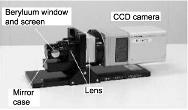
Fig.8. High resolution image detector
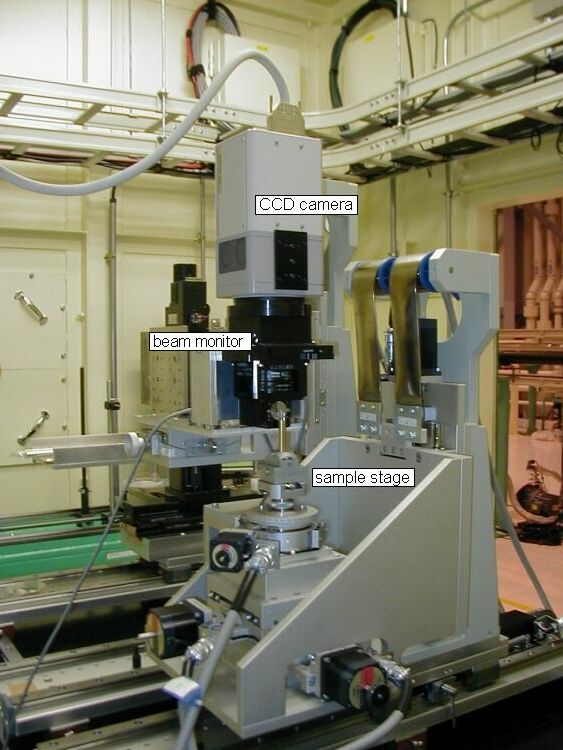
- Fig.9. High resolution X-ray tomographic system using high resolution 2-dimensional detector in experimental hutch 1
- Property of cooled CCD cameras
C4880-10-14A
(HAMAMATSU PHOTONICS)C4742-95HR
(HAMAMATSU PHOTONICS)pixel size 12 µm 5.9 µm CCD format 1000 (H) × 1018 (V) 4000 (H) × 2624 (V) dynamic range 14 bits 12 bits readout time 4 sec/frame 0.6 sec/frame