Microstructural imaging of cortical bone using monochromatic synchrotron radiation CT


Inquiry number
SOL-0000000977
Beamline
BL20B2 (Medical and Imaging I)
Scientific keywords
| A. Sample category | biology, medicine |
|---|---|
| B. Sample category (detail) | organism, cell, biological material |
| C. Technique | absorption and its secondary process |
| D. Technique (detail) | |
| E. Particular condition | 3D imaging (cf. CT), room temperature |
| F. Photon energy | X-ray (4-40 keV) |
| G. Target information | local structure, morphology |
Industrial keywords
| level 1---Application area | others |
|---|---|
| level 2---Target | |
| level 3---Target (detail) | organism |
| level 4---Obtainable information | density |
| level 5---Technique | imaging |
Classification
M60.20 X-ray CT
Body text
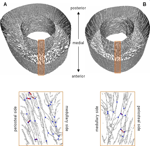
Synchrotron radiation combined with CT (SRCT) has opened up new possibilities in 3D microstructural analyses. The SRCT properties of natural collimation and extremely high light intensity allows the reconstruction of the highly resolved 3D cortical bone image. Furthermore, the availability of monochromatic X-rays eliminates beam-hardening artifacts and therefore allows image quantification for determining local bone mineralization. Figure shows the volume-rendered images of a pair of rat tibiae: normal(A) and subjected to sciatic neurectomy. Disuse-mediated canal network rarefaction as well as bone atrophy was clearly demonstrated, implying the involvement of reduced blood flow in bone atrophy.
[ T. Matsumoto, M. Yoshino, T. Asano, K. Uesugi, M. Todoh and M. Tanaka, Journal of Applied Physiology 100, 274-280 (2006), Fig. 4,
©2006 American Physiological Society ]
Source of the figure
Original paper/Journal article
Journal title
Journal of Applied Physiology, in press
Figure No.
Technique
Source of the figure
No figure
Required time for experimental setup
1 hour(s)
Instruments
| Instrument | Purpose | Performance |
|---|---|---|
| X-ray CT system | obtain 3-D internal structure | spatial resolution of about 10um |
References
| Document name |
|---|
| Matsumoto et al., Journal of Applied Physiology, in press |
Related experimental techniques
Questionnaire
The measurement was possible only in SPring-8. Impossible or very difficult in other facilities.
This solution is an application of a main instrument of the beamline.
Ease of measurement
Easy
Ease of analysis
Middle
How many shifts were needed for taking whole data in the figure?
Two-three shifts