Angiographic observation of tumor growth

問い合わせ番号
SOL-0000001145
ビームライン
BL20B2(医学・イメージングI)
学術利用キーワード
| A. 試料 | 生物・医学 |
|---|---|
| B. 試料詳細 | 生体(in vivo) |
| C. 手法 | 吸収、及びその二次過程 |
| D. 手法の詳細 | |
| E. 付加的測定条件 | 二次元画像計測, 時分割(ミリ秒) |
| F. エネルギー領域 | X線(4~40 keV) |
| G. 目的・欲しい情報 | 形態・巨視的構造 |
産業利用キーワード
| 階層1 | 製薬, その他 |
|---|---|
| 階層2 | 製剤 |
| 階層3 | 薬物, 生体 |
| 階層4 | 形態 |
| 階層5 | イメージング |
分類
A80.90 その他
利用事例本文
Microangiography with spatial resolution in the micrometer range has been carried out for depicting the microangioarchitecture of tumor-derived angiogenic vessels in a rabbit model of cancer using a high-resolution detector. A VX2 carcinoma was transplanted into a rabbit auricle. Small tumor blood vessels in an immature vascular network produced by angiogenesis were visualized after contrast agent injection. Images at 1, 3 and 7 days after transplantation are shown in Fig. 1. The tumor blood vessels exhibit a characteristic appearance, and small blood vessels of 20-30 µm diameter are depicted in these images.
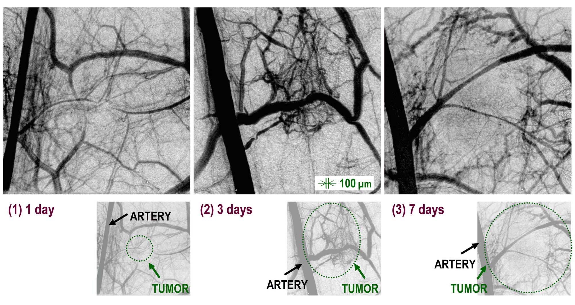
Fig. 1 Angiographic images of tumor blood vessels at 1, 3 and 7 d after tumor transplantation.
[ K. Umetani, T. Yamashita, N. Maehara and S. Imai, Journal of Institute of Image Information and Television Engineers 56, 492-494 (2002), Fig. 3,
©2002 The Institute of Image Information and Television Engineers ]
画像ファイルの出典
原著論文/解説記事
誌名
Journal of Institute of Image Information and Television Engineers 56(3) 492-494 (2002)
図番号
3
測定手法
The imaging system was composed of an X-ray direct-sensing type detector incorporating an X-ray SATICON pickup tube. Angiographic images were obtained and stored in a digital frame memory system with a 1024 x 1024-pixel, 10-bit format. Sequential images are obtained with an input field of 9.5 mm x 9.5 mm view and pixel size of 9.5 µm in the case of the 1024 x 1024-pixel format. Digital images are stored in 2 GB RAM of a specially designed frame memory system after analog-to-digital conversion. A personal computer system controls the entire imaging system including the camera system and the frame memory system.
画像ファイルの出典
図なし
測定準備に必要なおおよその時間
3 時間
測定装置
| 装置名 | 目的 | 性能 |
|---|---|---|
| Angiography system | Microangiography | 6-10 micron resolution |
参考文献
| 文献名 |
|---|
| 映像情報メディア学会誌 56(3) 492-494 (2002) |
関連する手法
no
アンケート
SPring-8だからできた測定。他の施設では不可能もしくは難しい
測定の難易度
中程度
データ解析の難易度
中程度
図に示した全てのデータを取るのにかかったシフト数
2~3シフト