単色放射光 CT による皮質骨微細構造イメージング


Inquiry number
SOL-0000001437
Beamline
BL20B2 (Medical and Imaging I)
Scientific keywords
| A. Sample category | biology, medicine |
|---|---|
| B. Sample category (detail) | organism, cell, biological material |
| C. Technique | absorption and its secondary process |
| D. Technique (detail) | |
| E. Particular condition | 3D imaging (cf. CT), room temperature |
| F. Photon energy | X-ray (4-40 keV) |
| G. Target information | local structure, morphology |
Industrial keywords
| level 1---Application area | others |
|---|---|
| level 2---Target | |
| level 3---Target (detail) | organism |
| level 4---Obtainable information | density |
| level 5---Technique | imaging |
Classification
M60.20 X-ray CT
Body text
単色放射光CTは非破壊で内部マイクロ構造を調べることのできる高精度な手法です。この手法を用いることで、今までは計測困難であった皮質骨内の脈管構造ネットワークの幾何学的特徴を測定することができます。骨内の脈管は骨に栄養を送る血管が走行する重要な構造です。さらに単色放射光を利用することで、ビームハードニングの影響を受けずに骨部分と脈管部の分離が容易となり、骨内のミネラル分布の定量化も可能となります。図に示すのは、ラット脛骨骨幹部の脈管構造に対する廃用の影響を測定した結果です。これより、廃用による骨萎縮とともに脈管ネットワークが退行することが明らかになり、廃用性骨萎縮(骨粗鬆症)には骨血流の低下が関与していることがわかりました。
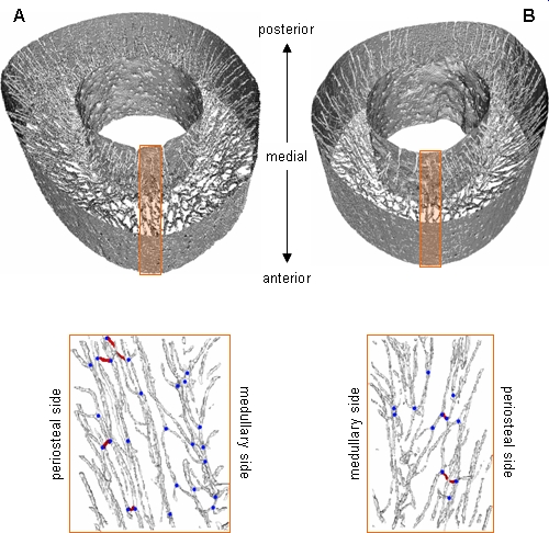
[ T. Matsumoto, M. Yoshino, T. Asano, K. Uesugi, M. Todoh and M. Tanaka, Journal of Applied Physiology 100, 274-280 (2006), Fig. 4,
©2006 American Physiological Society ]
Source of the figure
Original paper/Journal article
Journal title
Journal of Applied Physiology, in press
Figure No.
Technique
Source of the figure
No figure
Required time for experimental setup
1 hour(s)
Instruments
References
| Document name |
|---|
| Matsumoto et al., Journal of Applied Physiology, in press |
Related experimental techniques
Questionnaire
The measurement was possible only in SPring-8. Impossible or very difficult in other facilities.
This solution is an application of a main instrument of the beamline.
Ease of measurement
Easy
Ease of analysis
Middle
How many shifts were needed for taking whole data in the figure?
Two-three shifts