Nondestructive three-dimensional element concentration mapping by X-ray CT

Inquiry number
SOL-0000001107
Beamline
BL20B2 (Medical and Imaging I)
Scientific keywords
| A. Sample category | inorganic material |
|---|---|
| B. Sample category (detail) | insulator, ceramics, solid-state crystal, crystal |
| C. Technique | absorption and its secondary process |
| D. Technique (detail) | |
| E. Particular condition | 3D imaging (cf. CT) |
| F. Photon energy | X-ray (4-40 keV) |
| G. Target information | chemical state, crystal structure, dislocation, strain, elemental composition |
Industrial keywords
| level 1---Application area | industrial material, others |
|---|---|
| level 2---Target | Concrete |
| level 3---Target (detail) | |
| level 4---Obtainable information | crack, crevice, chemical state, structure, element distribution, molphology |
| level 5---Technique | imaging |
Classification
A80.30 inorganic material, A80.40 environmental materials, M60.20 X-ray CT
Body text
We succeeded in measuring the three-dimensional Cs concentration maps with high resolution under the nondestructive condition. This technique is based on the subtraction method and we acquired two set of X-ray CT images with two different photon energies just below and above the Cs-K absorption edge and obtained the differential images by subtraction. This subtraction method is known as a technique to obtain the qualitative information of the spatial distribution of elements. We performed some data corrections and made it possible to obtain quantitative maps of element concentration. Furthermore, by using the high spatial resolution X-ray CT system developed at BL20B2 of SPring-8, high spatial resolution of 20 m was achieved.
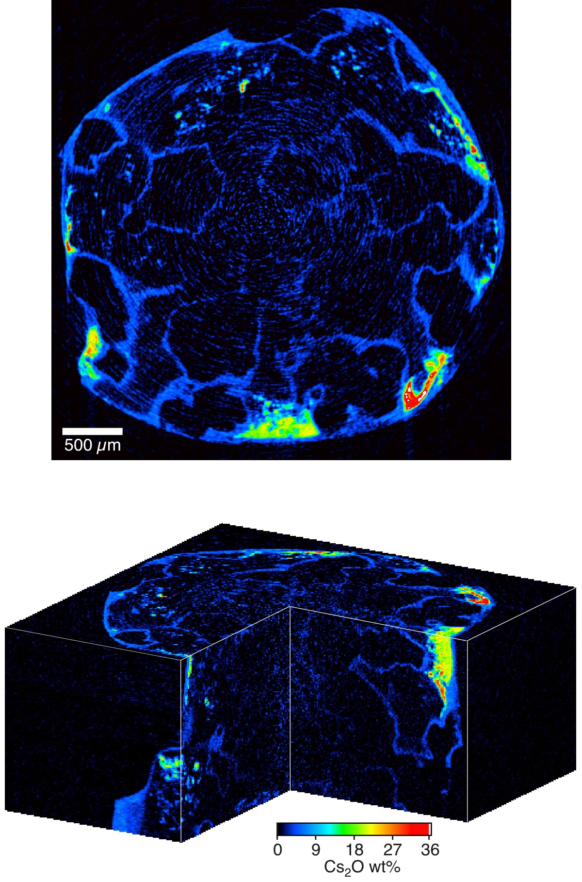
Two-dimensional and three-dimensional maps of Cs2O concentration in the Cs-doped partially molten granite sample
[ S. Ikeda, T. Nakano, A. Tsuchuyama, K. Uesugi, Y. Suzuki, K. Nakamura, Y. Nakashima and H. Yoshida, American Mineralogist 89, 1304-1313 (2004), Fig. 3(b), 3(f),
©2004 Mineralogical Society of America ]
Source of the figure
Original paper/Journal article
Journal title
S. Ikeda et al., Am. Mineral. 89, 1304 (2004)
Figure No.
Technique
Source of the figure
No figure
Required time for experimental setup
1 shift(s)
Instruments
| Instrument | Purpose | Performance |
|---|---|---|
| X-ray CT system | obtain internal structure of material | spatial resolution of 10um |
References
| Document name |
|---|
| S. Ikeda et al., Am. Mineral. 89, 1304 (2004) |
Related experimental techniques
Questionnaire
The measurement was possible only in SPring-8. Impossible or very difficult in other facilities.
This solution is an application of a main instrument of the beamline.
Ease of measurement
Middle
Ease of analysis
Middle
How many shifts were needed for taking whole data in the figure?
Two-three shifts