肺の屈折コントラストイメージング


Inquiry number
SOL-0000001447
Beamline
BL20B2 (Medical and Imaging I)
Scientific keywords
| A. Sample category | biology, medicine, research on method, instrumentation |
|---|---|
| B. Sample category (detail) | organic material, macromolecule, biology (in vivo), biology (in vitro), organism, cell, biological material, biomolecule, crystal, biomolecule, noncrystal |
| C. Technique | X-ray diffraction, absorption and its secondary process |
| D. Technique (detail) | reflection, refraction |
| E. Particular condition | 2D imaging, interface, room temperature |
| F. Photon energy | X-ray (4-40 keV), X-ray (> 40 keV) |
| G. Target information | morphology |
Industrial keywords
| level 1---Application area | Pharmaceuticals |
|---|---|
| level 2---Target | process analytical technology (PAT) |
| level 3---Target (detail) | drug, organism |
| level 4---Obtainable information | density, crack, crevice, molphology |
| level 5---Technique | imaging |
Classification
A80.50 Pharmaceuticals
Body text
PBI (Propagation-Based Imaging,屈折コントラストイメージングとも呼ばれる)は,物質中の屈折率の大きな変化を用いてX線のエッジ強調画像を作り出す手法です。生体組織では,特に組織と空気や骨との境界を明確に可視化します。下の画像は,生まれた直後のウサギ肺への空気の浸透を観察したものです。このように,PBI は生体中の空気を高分解能で観察するのに適した手法です。
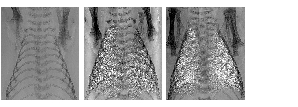
[ Physics in Medicine and Biology 50, 5031-5040 (2005), Fig.2, ©2005 Institute of Physics and IOP Publishing, Ltd. ]
Source of the figure
Original paper/Journal article
Journal title
R. Lewis et al., Physics in Medicine and Biology. 50 (2005) 5031-5040.
Figure No.
2
Technique
Source of the figure
No figure
Required time for experimental setup
1 shift(s)
Instruments
References
| Document name |
|---|
| R. Lewis et al., Physics in Medicine and Biology. 50 (2005) 5031-5040. |
Related experimental techniques
Questionnaire
The measurement was possible only in SPring-8. Impossible or very difficult in other facilities.
This solution is an application of a main instrument of the beamline.
Ease of measurement
Easy
Ease of analysis
Easy
How many shifts were needed for taking whole data in the figure?
Less than one shift