BL27SU B-branch
問い合わせ番号
INS-0000001445
B-branch: Higher energy soft X-ray spectroscopy and microscopy
The B-branch is dedicated for the higher-energy soft X-ray spectroscopy and microscopy analysis. A Si(111) double crystal monochromator provides the soft X-rays in the range 2.1-3.3 keV. The major applications of the B-branch are soft X-ray photoabsorption spectroscopy and scanning X-ray microscopy using soft X-ray micro beam under low-vacuum (〜1Pa) condition.
- Optics
The M0' in the optics hutch diverts the photon beam into B-branch. The main optical components are a Si (111) double-crystal monochromator and a K-B (Kirkpatrick and Baez) focusing system. Synchronous tuning between the undulator gap and the rotating angle of Si(111) monochromator maximizes the photon flux onto a sample. The Ni coated K-B focusing mirrors focuses the photon beam onto the sample, and the beam size was 15 (H) × 15 (V) µm2.
- X-rays at sample
Energy range 2.1 ∼ 3.3 keV Photon flux 1 × 10¹¹ photons/s/100 mA Beam size 15(H) × 15(V) µm - Experimental station
An experimental chamber installed on the B-branch offers a versatile soft X-ray application. The apparatus is used for the soft X-ray spectroscopy and microscopy experiments using monochromatized soft X-ray beam. The standard soft X-ray photoabsorption measurements are a total electron yield (TEY) and partial fluorescence yield (PFY) mode. TEY spectrum is obtained by a sample current method. For the PFY measurement, silicon drift-chamber detector (SDD) is prepared. Simultaneous detection of fluorescence and electron yield is possible for depth selective XAS analysis. If the sample thickness is thin enough for the soft X-ray transmission, transmission mode is also available. Standard pressure of analysis chamber is 1Pa.
Beam size at sample point is about 15 μm. The SDD detector is also available to the trace element analysis of light elements (i.e. XRF analysis) and elemental mapping analysis. Especially, the combination of XAS and XRF using a micro-focused X-ray beam allow determination of both the chemical state of environmentally important trace elements and their spatial distribution in biogenic carbonates [1].
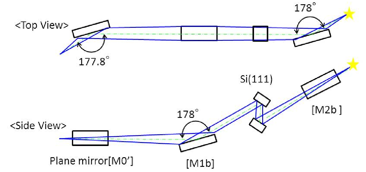
Layout of the optical components installed on the B-branch

Setup for the Soft X-ray photoabsorption measurement at B-branch.
- Standard equipment
- Energy dispersive soft X-ray analyzer (Amptek.:SDD Detector)
Effective area: 25mm2
Vacuum window: Si3N4 window or Be window
Sampling rate: <1×10⁵cps - Mapping stage
Available sample size 20mm×100mm(t1〜2)
- Reference
[1] Y. Tamenori et al., J. Structural Biol, 186, 214-223 (2014).