BL27SU C-branch
問い合わせ番号
INS-0000001446
C-branch: Lower energy soft X-ray spectroscopy
The C-branch is a high-resolution soft X-ray spectroscopy branch in the range 0.17-2.2 keV. The monochromator provides the intense photon beam with a photon bandwidth narrower than natural line width. The major applications of C-branch are soft X-ray photoabsorption spectroscopy and soft X-ray fluorescence/emission spectroscopy. The experimental stations can be connected to the beamline through the differential pumping apparatus or Si3N4 vacuum window. This setting makes us possible to control sample environments from 1 atom (helium) to 10-6 Pa and dedicated for in-situ and operando analysis.
- Monochromator and post-focusing mirrors
The monochromator is a varied-line-spacing plane grating (VLS-PGM) fixed deviation instrument of the Hettrick type. The monochromator consists of the entrance slit (S1), spherical mirrors (M21, M22), three gratings (G1, G2, G3), and exit slit (S2). Two spherical mirrors and three gratings allow the scanning of an energy range from 0.17 to 2.2 keV.
The re-focusing system is equipped with three types of optical elements, and has focusing points in tandem. At the two upstream stations (C1 and C2), toroidal mirror (M32) is used. At the most downstream station (C3), vertical and horizontal directions are focused by two sagittal-arranged cylindrical mirrors (M33, M4). Especially, the vertical direction of the photon beam is focused to 10 µm or less on the C3 station.
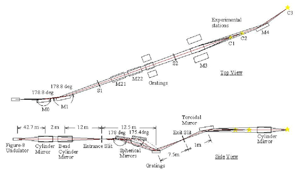
Layout of valid line-spacing plane grating monochromator
-
Parameters of spherical mirrors and gratings
Energy range (keV) 1.1 ~ 2.3 0.8 ∼ 2.3 0.4 ∼ 1.2 0.2 ∼ 0.6 Angle (deg) 88 88 86.7 86.7 Grating Grating name G1 Ni G1 Au G2 G3 Groove density 1200/mm/VLSPG 1200/mm/VLSPG 1400/mm/VLSPG 600/mm/VLSPG Material Ni/Si Au/Si Au/Si Au/Si Spherical mirror Mirror name M21 M21 M22 M22 Material Au/Si Au/Si Au/Si Au/Si -
Parameters of re-forcusing mirrors
Station C C2 C3 Mirror name M34 M32 M33 M4 Shape Plane Toroid Cylinder
(horizontal focus)Cylinder
(vertical focus)Incident angle (deg) 89 89 89 89 Material Au/Si Au/Si Au/Si Au/Si Beamsize V×H (µm) - 200 × 200 10 × 200
- X-rays at sample
Energy range 1st (Horizontal polarization) : 0.26 ∼ 2.2 keV
0.5th (Vertical polarization) : 0.17 ∼2 keVPhoton flux ∼ 1011 photon/s/100 mA/0.02%B.W. Energy resolution E/ΔE > 104
- Three experimental chambers are available.
Soft X-ray photoabsorption spectroscop
Normal ambient pressure soft X-ray photoabsorption spectroscopy
Soft-x-ray emission spectroscopy
- Soft X-ray spectroscopy
Energy dispersive soft X-ray analyzer (Amptek.:Silicon-Drift-Detector)
Soft X-ray emission spectrometer
Differential pumping system for the ambient atmospheric pressure experiments
[1] H. Ohashi, Nucl. Instr. Methods A467-468, 533 (2001).
[2] Y. Tamenori, J. Shynchrotron Rad.20, 419-425 (2013).
[3] Y. Tamenori, M. Morita, and T. Nakamura, J. Synchrotron Rad. 18, 747-752 (2011).
Soft X-ray photoabsorption analysis
This chamber offers versatile soft X-ray photoabsorption analysiss. The base pressure of the chamber is 5×10⁻⁶ Pa, therefore this apparatus is dedicated for the XAS measurements under high-vacuum condition. The standard soft X-ray photoabsorption measurements are a total electron yield (TEY) and partial fluorescence yield (PFY) mode. TEY spectrum is obtained by a sample current method. For the PFY measurement, silicon drift-chamber detector (SDD) was prepared[1]. Simultaneous detection of fluorescence and electron yield is possible for depth selective XAS analysis. Polarization dependence analysis is also possible by switching the direction of E-vector (vertical polarization and horizontal polarization) by selecting an appropriate undulator gap.
- Standard equipments
- Total electron yield obtained by sample current or metal anode detector
- Energy dispersive soft X-ray analyzer (SDD.:Silicon-Drift-Detector)
Effective area: 25 mm2
Si3N4 vacuum window, which can provides and soft X-ray down to the 300 eV (B Kα) - Air Lock
- Grove box is prepared for unexposed sample transfer to the analysis chamber
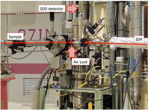
Overview of XAS analysis chamber
- Reference
[1] Y. Tamenori, M. Morita, and T. Nakamura, J. Synchrotron Rad. 18, 747-752 (2011).
Soft X-ray photoabsorption spectroscopy under the ambient atmospheric pressure condition
The standard procedure in soft X-ray experiments is to install all equipment necessary for experiments inside the high-vacuum chamber, since soft X-rays are fairly absorbed in atmospheric air. The high-vacuum chamber produces an ideally clean environment, therefore, it gives good opportunities to investigate a clean surface and anaerobic materials. On the other hand, the vacuum condition has drawbacks. Technologically significant phenomena take place under higher-pressure regions (1~760 mbar). One approach to overcoming the absorption in atmospheric air is to use a Helium-path instead of an air-path. Helium gas has a higher transmission than air in the soft X-ray region due to its smaller atomic number. In the BL27SU, a differential pumping system was developed for use with windowless soft X-ray experiments under normal atmospheric pressure helium conditions. This technique can eliminate the loss of incident photons due to the absorption of vacuum windows [1].
- Standard equipments
- Energy dispersive soft X-ray analyzer (Ourstex co. Ltd.:Silicon-Drift-Detector)*Currently out of use.
Effective area: 25 mm2
Si3N4 window, which can provides and soft X-ray down to the 0.3 keV (C Kα) - Differential pumping system for the ambient atmospheric pressure experiments
Four stage differential pump
Windowless connection to the beamline
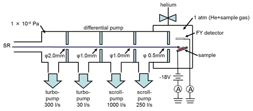
Layout of the differential pumping system for the ambient atmospheric pressure experiments
- Reference
[1] Y. Tamenori, J. Shynchrotron Rad., 20, 419-425 (2013).
Soft-x-ray emission spectroscopy
Electronic states of materials can be studied by means of soft x-ray emission spectroscopy (XES). The spot sizes at the sample position are ~ 10 and ~ 200 μm along the vertical and horizontal directions, respectively. The XES monochromator, which covers an energy range of 0.2 ~ 1.4 keV, is composed of a varied-line-spacing cylindrical grating and a CCD detector. The monochromator has no entrance slit to get a high efficiency. The energy resolution is about 0.4 eV when XES of 0.5 keV is detected and is about 1.5 eV at 1.0 keV. The SDD detector is also installed for the partial fluorescence yield XAS analysis with high detection efficiency. Therefore, both XAS and XES analysis is available.
- Standard equipments
- Energy dispersive soft X-ray analyzer (for partial-yield fluorescence yield measurements)
Effective area: 25 mm2
Si3N4 window, which can provides and soft X-ray down to the 0.3 keV (C Kα) - Soft X-ray emission spectrometer
Varied-line-spacing cylindrical grating and a CCD detector
Energy range : 0.2〜1.4 keV
Energy resolution :ΔE = 0.4 eV@500 eV、ΔE = 1.5 eV@1000 eV
- Energy dispersive soft X-ray analyzer (for partial-yield fluorescence yield measurements)
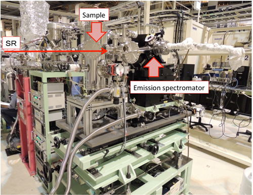
Overview of Soft X-ray emission spectromator