White X-ray topography combined with topo-tomographic technique

Inquiry number
SOL-0000001022
Beamline
BL28B2 (White Beam X-ray Diffraction)
Scientific keywords
| A. Sample category | inorganic material |
|---|---|
| B. Sample category (detail) | semiconductor, crystal |
| C. Technique | X-ray diffraction |
| D. Technique (detail) | |
| E. Particular condition | 3D imaging (cf. CT) |
| F. Photon energy | X-ray (> 40 keV) |
| G. Target information | dislocation, strain |
Industrial keywords
| level 1---Application area | Semiconductor |
|---|---|
| level 2---Target | silicon semiconductor |
| level 3---Target (detail) | SOI, substrate |
| level 4---Obtainable information | d-spacing (lattice parameter), structure |
| level 5---Technique | imaging |
Classification
A80.12 semiconductor, M10.10 single crystal diffraction
Body text
The advantage of the white x-ray topography combined with topo-tomographic technique is the ability to acquire information about the configuration of the dislocations from the variation in their features observed in a specific Laue spot by the tomographic technique, in addition to an information about the image contrast of the dislocations observed in several Laue spots by conventional white X-ray topography.
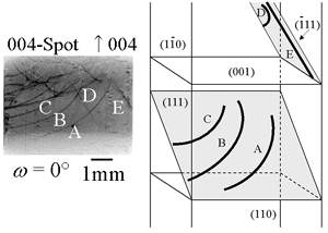
Fig. Topograph of CZ-silicon and schematic illustration showing results of identification of observed dislocations A to E.
[ S. Kawado, T. Taishi, S. Iida, Y. Suzuki, Y. Chikaura, K. Kajiwara, Journal of Synchrotron Radiation 11, 304-308 (2004), Fig. 4, 6,
©2004 International Union of Crystallography ]
Source of the figure
Original paper/Journal article
Journal title
S. Kawado, T. Taishi, S. Iida, Y. Suzuki, Y. Chikaura and K. Kajiwara J. Synchrotron Rad. (2004). 11, 304-308
Figure No.
4
Technique
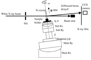
Fig.1 Schematic illustration of experimental arrangement.
[ S. Kawado, T. Taishi, S. Iida, Y. Suzuki, Y. Chikaura and K. Kajiwara, Journal of Synchrotron Radiation 11, 304-308 (2004), Fig. 1,
©2004 International Union of Crystallography ]
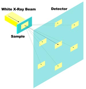
Fig.2 White X-ray Topography.
Source of the figure
Original paper/Journal article
Journal title
S. Kawado, T. Taishi, S. Iida, Y. Suzuki, Y. Chikaura and K. Kajiwara J. Synchrotron Rad. (2004). 11, 304-308
Figure No.
1
Required time for experimental setup
8 hour(s)
Instruments
References
Related experimental techniques
Questionnaire
The measurement was possible only in SPring-8. Impossible or very difficult in other facilities.
Ease of measurement
With a great skill
Ease of analysis
Middle
How many shifts were needed for taking whole data in the figure?
Four-nine shifts