X-ray diffraction microscopy
Inquiry number
SOL-0000000970
Beamline
BL29XU (RIKEN Coherent X-ray Optics)
Scientific keywords
| A. Sample category | inorganic material, organic material, biology, medicine |
|---|---|
| B. Sample category (detail) | metal, alloy, semiconductor, insulator, ceramics, solid-state crystal, amorphous, glass, organism, cell, biological material |
| C. Technique | X-ray diffraction |
| D. Technique (detail) | coherent scattering, phase measurement, speckle |
| E. Particular condition | 2D imaging, 3D imaging (cf. CT), X-ray microscopy |
| F. Photon energy | X-ray (4-40 keV) |
| G. Target information | structure analysis |
Industrial keywords
| level 1---Application area | Pharmaceuticals |
|---|---|
| level 2---Target | drug design, food, Environmental material |
| level 3---Target (detail) | organism |
| level 4---Obtainable information | |
| level 5---Technique | diffraction, imaging |
Classification
A80.12 semiconductor, A80.30 inorganic material, A80.32 organic material, A80.90 others
Body text
X-ray diffraction microscopy is a unique technique to reconstruct a sample image from its oversampled diffraction intensity pattern. Using this technique, one can determine electron density distribution without need of sample crystallization.
The following figures show the diffraction intensity pattern and the 2D reconstruction of Escherichia coli bacteria.
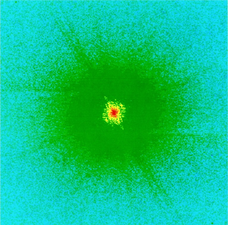
Fig. X-ray diffraction intensity pattern of Escherichia coli bacteria
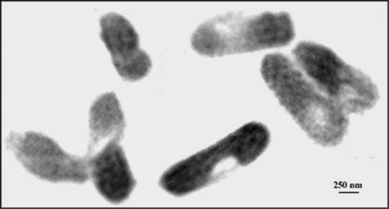
Fig. Image of Escherichia coli bacteria reconstructed from the diffraction intensity pattern
[ J. Miao, K. O. Hodgson, T. Ishikawa, C. A. Larabell, M. A. LeGros and Y. Nishino, Proceedings of National Academy of Science of the USA 100, 110-112 (2003), Fig. 1A, 2,
©2003 National Academy of Science ]
Source of the figure
Original paper/Journal article
Journal title
J. Miao, K.O. Hodgson, T. Ishikawa, C.A. Larabell, M.A. LeGros, and Y. Nishino, Proc. Natl. Acad. Sci. USA 100, 110 (2003)
Figure No.
1A,2
Technique
A sample is illuminated by coherent x-rays and its Fraunhofer diffraction intensities are measured using a two-dimensional x-ray detector.
Source of the figure
No figure
Required time for experimental setup
24 hour(s)
Instruments
| Instrument | Purpose | Performance |
|---|---|---|
| x-ray diffraction microscope |
References
| Document name |
|---|
| J. Miao, Y. Nishino, Y. Kohmura, B. Johnson, C. Song, S.H. Risbud, and T. Ishikawa, Phys. Rev. Lett. 95, 085503 (2005) |
| Y. Nishino, J. Miao, and T. Ishikawa, Phys. Rev. B 68, 220101(R) (2003) |
| J. Miao, J.E. Amonette, Y. Nishino, T.I shikawa, and K.O. Hodgson, Phys. Rev. B 68, 012201 (2003) |
| J. Miao, T.Ishikawa, E.H. Anderson, and K.O. Hodgson, Phys. Rev. B 67, 174104 (2003) |
| J. Miao, K.O. Hodgson, T. Ishikawa, C.A. Larabell, M.A. LeGros, and Y. Nishino, Proc. Natl. Acad. Sci. USA 100, 110 (2003) |
| J. Miao, T. Ishikawa, B. Johnson, E.H. Anderson, B. Lai, and K.O. Hodgson, Phys. Rev. Lett. 89, 088303 (2002) |
Related experimental techniques
Questionnaire
With user's own instruments.
Ease of measurement
With a great skill
Ease of analysis
With a great skill
How many shifts were needed for taking whole data in the figure?
Two-three shifts