走査型X線磁気顕微鏡


Inquiry number
SOL-0000001608
Beamline
BL39XU (X-ray Absorption and Emission Spectroscopy)
Scientific keywords
| A. Sample category | research on method, instrumentation |
|---|---|
| B. Sample category (detail) | magnetic material |
| C. Technique | absorption and its secondary process |
| D. Technique (detail) | XAFS, XANES, MCD, LD |
| E. Particular condition | polarization (circular), microbeam (1-10 µm), 2D imaging, magnetic field (< 2 T), room temperature |
| F. Photon energy | X-ray (4-40 keV) |
| G. Target information | spin/magnetism |
Industrial keywords
| level 1---Application area | storage device |
|---|---|
| level 2---Target | HD,MO |
| level 3---Target (detail) | magnetic layer, magnetic head, spin valve |
| level 4---Obtainable information | magnetic moment, magnetic anisotropy, interface magnetic structure |
| level 5---Technique | XAFS, XMCD, imaging |
Classification
A80.14 magnetic materials, M40.30 XMCD
Body text
走査型X線磁気顕微鏡は、試料に含まれる個々の磁性元素について2次元磁気画像計測を行うことのできるユニークな装置です。得られる画像の空間分解能はおよそ2 mです。この方法は、3d遷移金属元素、希土類元素、5d貴金属元素を含む磁性体試料に適用できます。また、試料上のm程度の大きさの領域について、X線磁気円二色性 (XMCD) スペクトルやXAFSスペクトルを測定することができます。
図に示すのは、CoCrPtB面内磁化膜について測定した2次元磁気画像です。あらかじめ縞状の磁気パターンが記録された試料を持ちいました。この例では、X線のエネルギーをPtのL3吸収端 (11.56 keV) に合わせ、試料中のPtの磁化の大きさや向きに応じた濃淡を観測しました。最小2.4 m幅の磁気パターンまで解像できています。
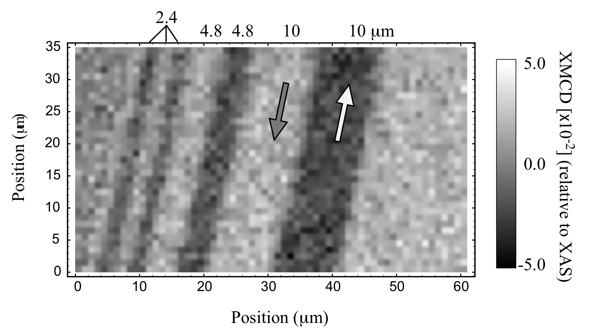
図. 走査型X線磁気顕微鏡で測定した、CoCrPtB面内磁化膜の2次元磁気画像
Source of the figure
Bulletin from SPring-8
Bulletin title
ナノテクノロジー総合支援プロジェクト研究成果報告書 Vol.4, 2004年A
Page
p. 147
Technique
走査型X線磁気顕微鏡は、ダイヤモンド移相子とX線用の集光ミラー (KBミラー) による光学系から成ります。移相子で生成した円偏光X線を、KBミラーを使って試料上で2 m 程度のスポットに集光します。円偏光の向きを切り替えながら試料からの蛍光X線強度をモニターすることで、ビーム位置での試料のX線磁気円二色性 (XMCD) 信号を測定します。このXMCD信号から、試料の磁化の大きさや磁化の向きに関する情報が得られます。試料の位置を精密ステージで動かすことで試料上でのX線ビーム位置を走査し、2次元画像を得ます。
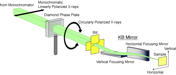
図. 走査型X線磁気顕微鏡の光学系
Source of the figure
Bulletin from SPring-8
Bulletin title
ナノテクノロジー総合支援プロジェクト研究成果報告書 Vol. 4, 2004年A
Page
p. 146
Required time for experimental setup
3 shift(s)
Instruments
| Instrument | Purpose | Performance |
|---|---|---|
| 走査型X線磁気顕微鏡 | 2次元磁気画像の測定、微小領域におけるXMCD測定 | 集光X線スポットサイズ 2ミクロン、光子数~10^10 photons/s |
References
| Document name |
|---|
| M. Takagaki, M. Suzuki, N. Kawamura, H. Mimura, and T. Ishikawa, The 8th International Conference on X-ray Microscopy (XRM2005), 26-30 July 2005, Himeji, Japan. |
Related experimental techniques
磁気力顕微鏡(MFM), 光電子顕微鏡 (PEEM), 透過型X線顕微鏡
Questionnaire
The measurement was possible only in SPring-8. Impossible or very difficult in other facilities.
This solution is an application of a main instrument of the beamline.
This solution is application of a new instrument installed in the past two years.
Ease of measurement
Easy
Ease of analysis
Easy
How many shifts were needed for taking whole data in the figure?
Two-three shifts