High energy MAD experiment using I and Xe

問い合わせ番号
SOL-0000001068
ビームライン
BL41XU(生体高分子結晶解析 I)
学術利用キーワード
| A. 試料 | 生物・医学, 計測法、装置に関する研究 |
|---|---|
| B. 試料詳細 | 結晶, 生体高分子、結晶, 蛋白質, 医薬品 |
| C. 手法 | X線回折, 吸収、及びその二次過程, 蛍光X線 |
| D. 手法の詳細 | 単結晶構造解析, MAD, XAFS |
| E. 付加的測定条件 | 低温(〜液体窒素) |
| F. エネルギー領域 | X線(4~40 keV) |
| G. 目的・欲しい情報 | 分子構造 |
産業利用キーワード
| 階層1 | 製薬 |
|---|---|
| 階層2 | ドラッグデザイン, 製剤 |
| 階層3 | タンパク質, 薬物 |
| 階層4 | 高次構造, 絶対構造決定 |
| 階層5 | XAFS, 回折 |
分類
A80.50 製薬, M10.10 単結晶回折, M40.10 XAFS
利用事例本文
Multiwavelength anomalous diffraction (MAD) method is the powerful method to obtain the phase information from one sample crystal composed of molecule with heavy atoms.
In this case, the MAD experiments at near energies of iodine K-absorption (33.17 keV) and xenon K-absorption (34.56 keV) were done using third harmonics of undulator on BL41XU.
Sample crystals were prepared from hen egg white lysozyme. Iodine-doped crystals were obtained by co-crystallization with sodium iodide, and xenon-doped crystals were made by putting its under high-pressure (3 MPa.) xenon gas for 15 minutes.
Precise energies of absorption edge of both iodide and xenon were decided by measurement of X-ray absorption spectra from each derivative crystal (Fig. 1). The MAD experiments were done at near energies decided absorption edge, and the phase information of the lysozyme crystal were able to determined from those successfully. Figure 2 shows the positions of iodine and xenon bound to lysozyme molecule.
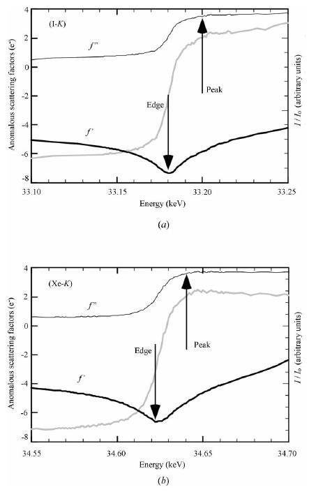
Figure 1. X-ray absorption spectra from (a) iodine and (b) xenon
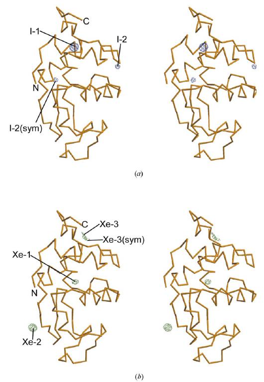
Figure 2. Bounding positions of (a) iodine and (b) xenon
[ K. Takeda, H. Miyatake, S.-Y. Park, M. Kawamoto, N. Kamiya, K. Miki, Journal of Applied Crystallography 37, 925-933 (2004), Fig. 2, 5,
©2004 International Union of Crystallography ]
画像ファイルの出典
原著論文/解説記事
誌名
K. Takeda, et al, J. Appl. Crystallogr. 37, 925-933 (2004)
図番号
2, 5
測定手法
Multiwavelength anomalous dispersion (MAD) method is that the X-ray diffraction data are collected at near several energies of the absorption edge of heavy atom binding to molecule consisting crystal. By comparing and analyzing those diffraction data, the phase information is decided, and three-dimensional structure is able to determined.
画像ファイルの出典
図なし
測定準備に必要なおおよその時間
1 時間
測定装置
| 装置名 | 目的 | 性能 |
|---|---|---|
| Protein Crystal Diffractometer | To record diffraction data | |
| Peltier-cooled Si-PIN photodiode | To measure X-ray absorption spectrum |
参考文献
| 文献名 |
|---|
| K. Takeda, et al. J. Appl. Crystallogr. 37, 925-933 (2004) |
関連する手法
アンケート
SPring-8だからできた測定。他の施設では不可能もしくは難しい
本ビームラインの主力装置を使っている
測定の難易度
熟練が必要
データ解析の難易度
中程度
図に示した全てのデータを取るのにかかったシフト数
2~3シフト