Ultra-high resolution structure analysis of endopolygaracturonase from C. purpureum

Inquiry number
SOL-0000001166
Beamline
BL41XU (Macromolecular Crystallography I)
Scientific keywords
| A. Sample category | atom, molecule, radical, biology, medicine |
|---|---|
| B. Sample category (detail) | biomolecule, crystal, protein, pharmaceuticals |
| C. Technique | X-ray diffraction |
| D. Technique (detail) | single crystal |
| E. Particular condition | low-T (~ liquid N2) |
| F. Photon energy | X-ray (4-40 keV) |
| G. Target information | chemical state, molecular structure, structure analysis, function and structure, charge density |
Industrial keywords
| level 1---Application area | environment, Pharmaceuticals |
|---|---|
| level 2---Target | drug design, process analytical technology (PAT), food |
| level 3---Target (detail) | protein, drug |
| level 4---Obtainable information | interatomic distance, crystal structure, local structure, electronic state, chemical state, absolute configuration |
| level 5---Technique | diffraction, X-ray diffraction |
Classification
A80.50 Pharmaceuticals, M10.10 single crystal diffraction
Body text
Endopolygalacturonases (endoPGs) catalyze random hydrolysis of the -1,4 glycosidic linkages in polygalacturonic acid, a component of pectin that is a major component of plant cell wall.
In the paper written in 2002 (Biochemistry, 41, 6651-6659, 2002), three-dimensional structure of this enzyme was determined at 0.96 Å using SPring-8/BL44B2 (Fig. 1).
To analyze structure of this enzyme at higher resolution, the conditions of crystallization and X-ray diffraction experiment were refined. The diffraction spots up to 0.76 Å resolution were recorded (Fig. 2), and an atomic structure was analyzed at 0.85 Å resolution using BL41XU (Fig. 3).
Currently, the diffraction data over 0.7 Å were collected with a large area detector and short wavelength (< 0.6 Å) X-ray derived from undulator third-harmonics.
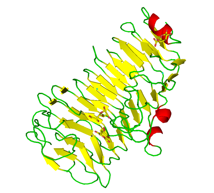
Figure 1. Schematic drawing of endoPG from C. purupureum (PDB ID: 1K5C)
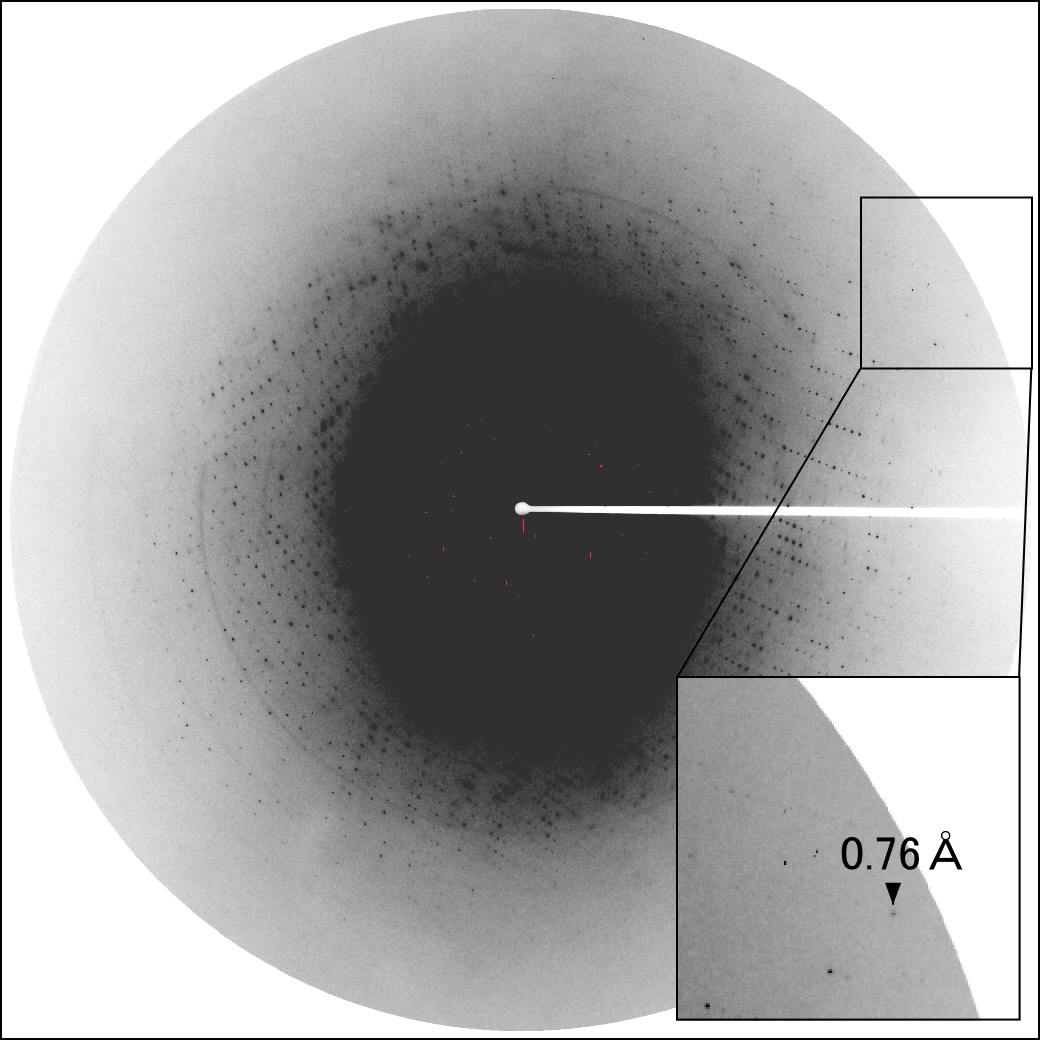
Figure 2. X-ray diffraction image of endoPG
This image was recorded using X-ray CCD detector (marCCD165) used at BL41XU in past.
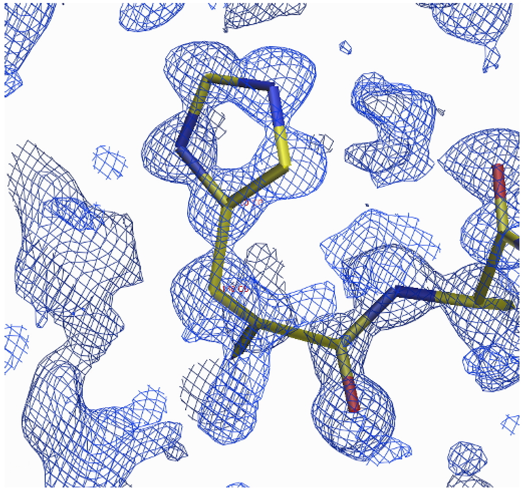
Figure 3. Electron density map of endoPG at 0.85 Å resolution
Source of the figure
Private communication/others
Description
コメント参照のこと
Technique
Source of the figure
No figure
Required time for experimental setup
8 hour(s)
Instruments
| Instrument | Purpose | Performance |
|---|---|---|
| Protein Crystal Diffractometer | To record diffraction data |
References
| Document name |
|---|
| T. Shimizu, et al., Biochemistry, 41, 6651-6659 (2002) |
Related experimental techniques
Questionnaire
The measurement was possible only in SPring-8. Impossible or very difficult in other facilities.
This solution is an application of a main instrument of the beamline.
Ease of measurement
With a great skill
Ease of analysis
Middle
How many shifts were needed for taking whole data in the figure?
Four-nine shifts