Recording of X-ray diffraction image from crystal with large unit cell

Inquiry number
SOL-0000001187
Beamline
BL41XU (Macromolecular Crystallography I)
Scientific keywords
| A. Sample category | biology, medicine |
|---|---|
| B. Sample category (detail) | biomolecule, crystal, protein, pharmaceuticals |
| C. Technique | X-ray diffraction |
| D. Technique (detail) | single crystal |
| E. Particular condition | low-T (~ liquid N2) |
| F. Photon energy | X-ray (4-40 keV) |
| G. Target information | local structure, structure analysis, crystal structure, morphology |
Industrial keywords
| level 1---Application area | Pharmaceuticals |
|---|---|
| level 2---Target | drug design, process analytical technology (PAT) |
| level 3---Target (detail) | protein, drug |
| level 4---Obtainable information | film thickness, d-spacing (lattice parameter), crystal structure, supra-molecular assemblies |
| level 5---Technique | diffraction |
Classification
A80.50 Pharmaceuticals, M10.10 single crystal diffraction
Body text
Some protein crystals have a large unit-cell over several hundred angstrom depending on the molecular weight, molecular numbers in an asymmetric unit or space group of crystal. These crystals can diffract X-ray weakly because that diffraction intensity is in inverse proportion to the volume of the crystal. Moreover, it is difficult to separate each diffraction spot on the detector.
In this case, it is introduced that the recording of X-ray diffraction image from the crystal with large unit-cell of 590 Å.
This image was collected from crystal of calcium ATPase from rat sarcoplasmic reticulum. The crystal was soaked to aurothioglucose solution as a contrast medium to visualize the lipid bilayer in it.
Using high brilliant and high parallel beam from undulator of BL41XU, each diffraction spots were separated clearly on the detector surface (Fig. 1).
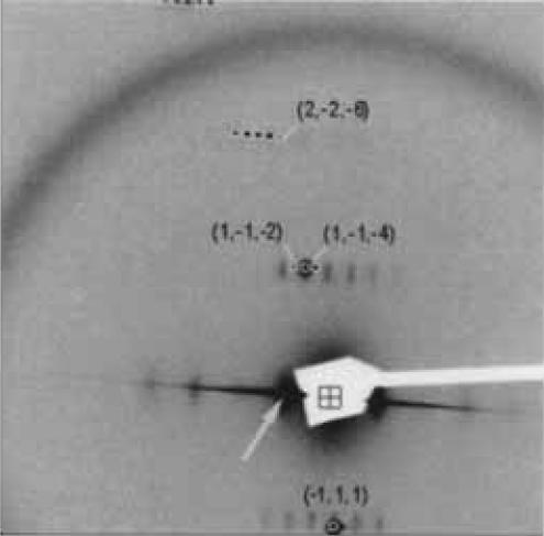
Figure 1. X-ray diffraction image of calcium ATPase from rat sarcoplasmic reticulum
Space group and unit cell of the crystal is P41 and a=b=71.7, c=590.3 Å.
Using wavelength is 1.0401 Å, and the used detector is RIGAKU R-AXIS V with 600 mm distance.
Source of the figure
Bulletin from SPring-8
Bulletin title
C. Toyoshima and T. Tsuda, SPring-8 User Experimental Report 12, 212 (2003B)
Page
212
Technique
Source of the figure
No figure
Required time for experimental setup
8 hour(s)
Instruments
| Instrument | Purpose | Performance |
|---|---|---|
| Protein Crystal Diffractometer | To record diffraction data | |
| Protein Crystal Diffractometer | To record diffraction image | sensitive area: 400mm x 400mm |
References
| Document name |
|---|
| C. Toyoshima and T. Tsuda, SPring-8 User Experimental Report 12, 212 (2003B) |
Related experimental techniques
Questionnaire
The measurement was possible only in SPring-8. Impossible or very difficult in other facilities.
This solution is an application of a main instrument of the beamline.
With user's own instruments.
Ease of measurement
With a great skill
Ease of analysis
With a great skill
How many shifts were needed for taking whole data in the figure?
Two-three shifts