BL44XU OUTLINE
Inquiry number
INS-0000000389
ABSTRACT
This beamline is specially designed to collect high quality X-ray diffraction data of biological macromolecular assemblies, e.g. protein complexes, protein-nucleic acid complexes, and viruses.
AREA OF RESEARCH
- Crystal structure analysis of biological macromolecular assemblies (e.g. membrane protein complexes, protein complexes, protein-nucleic acid complexes, and viruses)
KEYWORDS
- Scientific field
Biological macromolecular assemblies, Protein crystallography, Structural biology - Equipment
High precision goniometer, Imaging plate detector, CCD detector, Cryostream cooler
SOURCE AND OPTICS
Main beamline optics is a double-crystal monochromator and a horizontal focusing mirror.
| Insertion device | In-vacuum undulator |
| Undulator period : λu | 32 mm |
| Number of period : Nperiod | 140 |
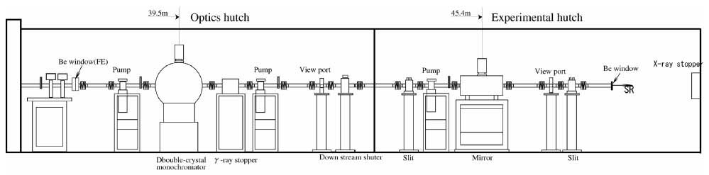
Schematic View of Beamline
- X-rays at sample
Energy (wavelength) Range 6.5 ~17.7 keV (1.9 Å〜0.7 Å) Energy Resolution ΔE/E < 2 × 10⁻⁴ Photon Flux 3.0 × 10¹² photons/s (φ50 µm) Beam Size φ70 µm, φ50 µm, φ30 µm, φ20 µm, φ10 µm
EXPERIMENTAL STATION
- Goniometer
SMARGON (SmarAct GmbH)Rotation axis range ω: -1270 〜 1270°
χ: -0.5 〜 30°
φ: -1270 〜 1270°
Deviation from rotation axis ω: < 1 μm
χ: < 7 μm
φ: < 7 μm
Rotation speed ω: <180 °/sec
χ: < 10°/sec
φ: < 80°/sec
Collimation Pinholes Sample camera Co-axial CCD camera - Cryostream cooler
Temperature range 90 ∼ 300 K (N2 gas)
35 ∼ 300 K (He gas)Nozzle inside diameter 7mmφ - Sample changer
SPACE-II - Detector
Eiger X 16M (Dectris)Number of detector modules 4 x 8 Active area (width x height) 311.2 x 327.8 mm2 Pixel size 75 x 75 µm2 Inactive area 6.6 % PSF 1 pixel Maximum frame rate 133 Hz Maximum count rate 5 x 10⁸ photons/s/mm2 Counter bit depth 12 bit Image bit depth 16 or 32 bit Data format HDF5 / NeXus - Main data processing, structure determination software
- XDS
- MOSFLM
- autoPROC
- CCP4
| Sample to detector distance | 115 – 1200 mm |
| Offset (vertical) | 0 – 150 mm |
| 2θ | 0 – 15° |
PUBLICATION SEARCH
* Sorry, Some parts of results are displayed using Japanese characters.
CONTACT INFORMATION
Eiki YAMASHITA
Research Center for Structural and Functional Proteomics
Institute for Protein Research, Osaka University
3-2 Yamadaoka, Suita, Osaka, 565-0871
Phone : +81-(0)6-6879-8635
Fax : +81-(0)6-6879-4313
@ SPring-8
Phone: 0791-58-1814
Fax : 0791-58-1814
e-mail : 
Website
http://www.protein.osaka-u.ac.jp/rcsfp/supracryst/en/research/beamline/