単一筋原繊維からの回折

Inquiry number
SOL-0000003405
Beamline
BL45XU (Macromolecular Crystallography II)
Scientific keywords
| A. Sample category | biology, medicine, research on method, instrumentation |
|---|---|
| B. Sample category (detail) | organism, cell, protein |
| C. Technique | X-ray diffraction |
| D. Technique (detail) | small angle scattering |
| E. Particular condition | microbeam (1-10 µm), 2D imaging |
| F. Photon energy | X-ray (4-40 keV) |
| G. Target information | structure analysis, structural hierarchy |
Industrial keywords
| level 1---Application area | Pharmaceuticals, others |
|---|---|
| level 2---Target | fiber |
| level 3---Target (detail) | protein, organism |
| level 4---Obtainable information | crystal structure, orientation (preferred orientation), structure |
| level 5---Technique | diffraction, X-ray diffraction, SAX, scattering, SAXS |
Classification
M20.10 SAX
Body text
筋肉細胞内にある直径わずか2マイクロメートルの筋原繊維1本からX線回折像を記録し、その中の収縮蛋白の格子構造を直接可視化することに成功した。これは、細胞内にある水和した蛋白質の集合体でX線回折像が記録された最小の例となる(体積にして 従来の約1/1000)。さらに蛋白集合体の単一格子からの回折像記録も世界初である。今回の成果は、他にも細胞内に多種存在する微小な機能性蛋白質集合体にX線回折による構造解析の道を拓くことになり、ポスゲノム時代の生物学に新たな研究手段を提供することになろう。
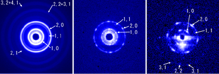
図:マルハナバチ飛翔筋筋細胞から実際に記録されたエンドオン回折像
(左はビームサイズ50μm、右の2つは2μm)。
Source of the figure
Bulletin from SPring-8
Bulletin title
http://www.spring8.or.jp/j/press/020806/muscle_fibril.html
Page
Technique
Source of the figure
No figure
Required time for experimental setup
6 shift(s)
Instruments
| Instrument | Purpose | Performance |
|---|---|---|
| BL45XU-SAXS | SAXS, Microbeam |
References
| Document name |
|---|
| Biophysical Journal Volume 83 August 2002 1074–1081 |
Related experimental techniques
Questionnaire
The measurement was possible only in SPring-8. Impossible or very difficult in other facilities.
With user's own instruments.
Ease of measurement
With a great skill
Ease of analysis
With a great skill
How many shifts were needed for taking whole data in the figure?
Four-nine shifts