X-ray microbeam diffraction (X-ray micro-diffraction)

Inquiry number
SOL-0000000927
Beamline
BL47XU (Micro-CT)
Scientific keywords
| A. Sample category | organic material, atom, molecule, radical, biology, medicine, research on method, instrumentation |
|---|---|
| B. Sample category (detail) | macromolecule, crystal, solution, liquid crystal, lipid, membrane, neutral molecule, biomolecule, crystal, protein, pharmaceuticals |
| C. Technique | X-ray diffraction |
| D. Technique (detail) | single crystal, powder diffraction, small angle scattering |
| E. Particular condition | microbeam (sub-µm), 2D imaging, tensile loading |
| F. Photon energy | X-ray (4-40 keV) |
| G. Target information | molecular structure, structure analysis, crystal structure, dislocation, strain, structural change |
Industrial keywords
| level 1---Application area | environment, Pharmaceuticals |
|---|---|
| level 2---Target | drug design, process analytical technology (PAT), fiber |
| level 3---Target (detail) | protein, drug, tablet |
| level 4---Obtainable information | crystal structure, orientation (preferred orientation), crystallinity, polymorphism, structure |
| level 5---Technique | diffraction, X-ray diffraction, SAX, imaging |
Classification
A60.20 environment, A80.32 organic material, A80.40 environmental materials, A80.50 Pharmaceuticals, M10.10 single crystal diffraction, M10.20 powder diffraction, M10.30 surface・interface diffraction, M20.10 SAX
Body text
X-ray microbeam diffraction (x-ray micro-diffraction) is an accurate technique to study crystal structures of very minute area with sub-micrometer level. Using this technique, one can measure molecular structures in a selected area of sample. Comparing with conventional x-ray diffraction, this technique has other following advantages;
- selectable exposure point
- available for very small crystal
- low background level
- reduce x-ray damage by moving the probe during exposure.
The figure shows the series of x-ray micro-diffraction diagrams of a fiber of high-strength biodegradable polyesters (poly[R)-3-hydroxybutyrate], 50 micron diameter) scanned perpendicular to the fiber axis with a step of 2 microns. Red arrows indicate a new reflection derived from different structure from other parts. These data reveal the fact that this fiber has core-and-sheath structure, with only helix conformation in sheath region and with both planar-zigzag conformation and helix conformation in core region.
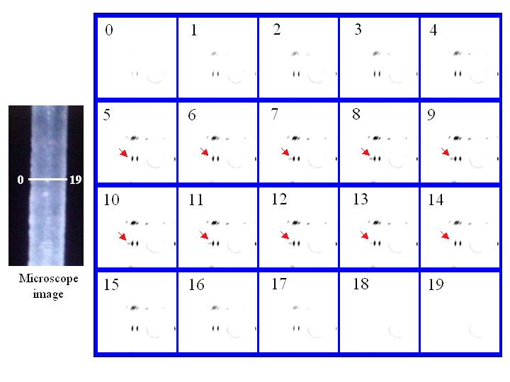
Figure: X-ray micro-diffraction diagrams of fiber of biodegradable polyesters (poly[R)-3-hydroxybutyrate], 50 micron diameter) recorded from the line area in microscope image (left picture).
[ T. Iwata, Y. Aoyagi, M. Fujita, H. Yamane, Y. Doi, Y. Suzuki, A. Takeuchi and K. Ursugi, Macromolecular Rapid Communications 25, 1100-1104 (2004), Fig. 3,
©2004 Wiley VCH ]
Source of the figure
Private communication/others
Description
理化学研究所 土肥高分子化学研究室の岩田忠久博士
Technique
Micro-diffraction experiment is performed by using a micro-focused beam generated with a Fresnel zone plate optic. In this solution, diffraction pattern from a small portion (~sub-micrometer) of a sample can be obtained.
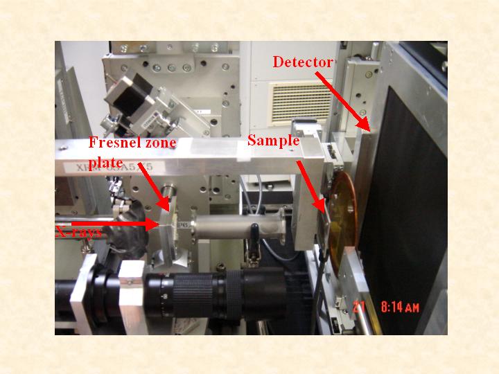
Experimental setup for x-ray micro-diffraction.
Source of the figure
Private communication/others
Description
理化学研究所 岩田忠久博士
Required time for experimental setup
1 day(s)
Instruments
| Instrument | Purpose | Performance |
|---|---|---|
| Fresnel zone plate | x-ray micro-focusing | 0.25 micron theoretical resolution |
| Beam monitor 2 | optical alignment | 4.3 micron pixel size |
| Image intensifier | detector | 4 inch field of view |
References
| Document name |
|---|
| T. Iwata et. al., Macromol. Rapid Commun. 25, 1100-1104, 2004 |
Related experimental techniques
Questionnaire
The measurement was possible only in SPring-8. Impossible or very difficult in other facilities.
Ease of measurement
Middle
Ease of analysis
Middle
How many shifts were needed for taking whole data in the figure?
Two-three shifts