Observation of microscopic voids of the concrete.


Inquiry number
SOL-0000001102
Beamline
BL47XU (Micro-CT)
Scientific keywords
| A. Sample category | inorganic material |
|---|---|
| B. Sample category (detail) | insulator, ceramics, solid-state crystal |
| C. Technique | absorption and its secondary process |
| D. Technique (detail) | |
| E. Particular condition | 3D imaging (cf. CT), room temperature |
| F. Photon energy | X-ray (4-40 keV) |
| G. Target information | dislocation, strain, morphology |
Industrial keywords
| level 1---Application area | construction, industrial material |
|---|---|
| level 2---Target | Concrete |
| level 3---Target (detail) | |
| level 4---Obtainable information | density, crack, crevice, structure |
| level 5---Technique | imaging |
Classification
A80.20 metal ・material, A80.30 inorganic material, M60.20 X-ray CT
Body text
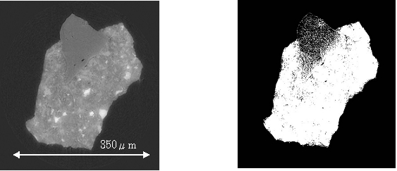
X ray CT is unique technique to study void structure of concrete. Concrete contains aggregates and cement paste. Cement paste is porous, and its voids are estimated as path of chemical components which causes concrete deterioration. X ray CT is enables of nondestructive observations of void structure of cement paste in three dimensions. Figure 1 shows cross section of ordinary Portland cement paste. Black parts are voids. In upper left side we can see sand. To extract voids part, cross sections are digitized. Figure 2 shows digitized cross section. With image processing data, we can estimate physical quantities as pore size distributions.
[ T. Hitomi, Y. Mita, H. Saito and N. Takeda, Proceedings of the Japan Concrete Institute 26, 645-650 (2004), Fig. 5, 10,
©2004 Japan Concrete Institute ]
Source of the figure
Original paper/Journal article
Journal title
SPring-8におけるX線CT像によるモルタル微細構造の観察,コンクリート工学年次論文集,Vol.26,pp645-650.2004.7
Figure No.
Technique
Source of the figure
No figure
Required time for experimental setup
1 shift(s)
Instruments
| Instrument | Purpose | Performance |
|---|---|---|
| X-ray CT system | obtain interlnal structure of materials | spatial resolution of about 1µm |
References
| Document name |
|---|
| SPring-8におけるX線CT像によるモルタル微細構造の観察,コンクリート工学年次論文集,Vol.26,pp645-650.2004.7 |
Related experimental techniques
Questionnaire
The measurement was possible only in SPring-8. Impossible or very difficult in other facilities.
This solution is an application of a main instrument of the beamline.
Ease of measurement
Easy
Ease of analysis
Middle
How many shifts were needed for taking whole data in the figure?
Less than one shift