高分解能結像型X線マイクロトモグラフィ

Inquiry number
SOL-0000001685
Beamline
BL47XU (Micro-CT)
Scientific keywords
| A. Sample category | inorganic material, organic material, biology, medicine, research on method, instrumentation |
|---|---|
| B. Sample category (detail) | metal, alloy, semiconductor, insulator, ceramics, amorphous, glass, organic material, macromolecule, biology (in vitro), organism, cell, biological material, biomolecule, noncrystal, pharmaceuticals, environmental material |
| C. Technique | absorption and its secondary process |
| D. Technique (detail) | |
| E. Particular condition | 3D imaging (cf. CT), X-ray microscopy |
| F. Photon energy | X-ray (4-40 keV) |
| G. Target information | morphology |
Industrial keywords
| level 1---Application area | Semiconductor, storage device, cell (battery), mechanics, construction, environment, Pharmaceuticals, industrial material |
|---|---|
| level 2---Target | silicon semiconductor, compound semiconductor, process analytical technology (PAT), fiber, Concrete |
| level 3---Target (detail) | wire, electric rod, drug, tablet |
| level 4---Obtainable information | density, crack, crevice, structure, molphology |
| level 5---Technique | imaging |
Classification
M60.20 X-ray CT
Body text
結像型X線マイクロトモグラフィは、X線CTの一種で、非常に高い空間分解能が特徴の高精度な手法です。この手法を用いることで、これまで観察が不可能だった1ミクロン以下の試料内部の3次元イメージを測定することができるようになりました。測定できる試料の大きさは、およそ直径100ミクロンまでです。
図に示すのは、珪藻土について本手法で測定した3次元再構成像です。この結果から、試料内の1ミクロン以下の微細な3次元構造が鮮明に見えていることがわかりました。
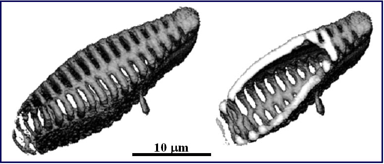
図 珪藻土のCT3次元像。左は全体像、右はデータ処理で試料を途中から切り落として表示したもの。
[ A. Takeuchi, K. Uesugi, H. Takano and Y. Suzuki, Review of Scientific Instruments 73, 4246-4249 (2002), Fig. 5,
©2002 American Institute of Physics ]
Source of the figure
Original paper/Journal article
Journal title
Rev. Sci. Instrum 73, 4246 (2002)
Figure No.
5
Technique
この実験では、高い分解能を得るために、X線像を、可視光顕微鏡と同じようにX線用のレンズ(フレネルゾーンプレートといいます)を使って拡大しています。そうすることによって、100nm程度までの細かい内部構造のイメージングを可能にしています。
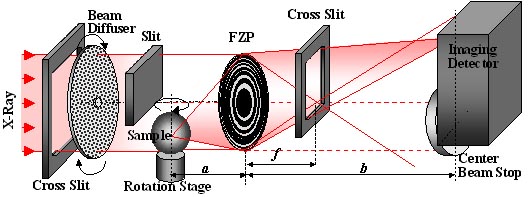
結像型X線マイクロトモグラフィのセットアップ図
Source of the figure
Private communication/others
Description
自作
Required time for experimental setup
2 day(s)
Instruments
| Instrument | Purpose | Performance |
|---|---|---|
| フレネルゾーンプレート | X線像の拡大 | 視野100ミクロン程度 |
| ビームモニタ2 | 光学系の調整、データ取得 | ピクセルサイズ4.3ミクロン |
| 精密回転ステージ | 試料の回転 | 回転軸ぶれ精度0.2ミクロン以下 |
References
| Document name |
|---|
| A. Takeuchi et. al., Rev. Sci. Instrum., 73, 4246-4249 (2002). |
Related experimental techniques
X線CT
Questionnaire
The measurement was possible only in SPring-8. Impossible or very difficult in other facilities.
This solution is an application of a main instrument of the beamline.
Ease of measurement
Middle
Ease of analysis
Middle
How many shifts were needed for taking whole data in the figure?
Less than one shift