Development of a New Type of X-Ray Microscopy for Three-Dimensional Observation of Cell Interior - Success in X-ray CT Scanning of Human Chromosomes for the First Time in the World (Press Release)
- Release Date
- 24 Dec, 2008
- BL29XU (RIKEN Coherent X-ray Optics)
Key research achievements
- Realization of a high-contrast X-ray imaging technology that enables the three-dimensional observation of the cell interior
- First observation of the axial structure of human chromosomes without any specified protein labeling
- Hope for medical application of the microscopy using X-ray free electron laser
RIKEN (Ryoji Noyori, President) developed a new type of X-ray microscopy that enables the three-dimensional high-contrast observation of the complete inner structure of cells for the first time in the world, and succeeded in observing the inner structure of human chromosomes. This is the achievement by Yoshinori Nishino, scientist, Yukio Takahashi, guest scientist (also, Special Lecturer of Osaka University), and Tetsuya Ishikawa, senior scientist of the Coherent X-Ray Optics Laboratory in RIKEN SPring-8 Center (Tetsuya Ishikawa, Director), and Kazuhiro Maeshima, scientist, and Naoko Imamoto, senior scientist of the Cellular Dynamics Laboratory of RIKEN Advanced Science Institute (Kohei Tamao, Director).
It has been difficult to observe the complete inner structures of cells and cell organelles by conventional microscopy. Cells and cell organelles of more than 1 μm thickness (μm: a millionth of a meter) are too thick to be observed by transmission electron microscopy using electrons. Although X-ray has a high level of penetrability and is suitable for the internal observation of thick materials, most of the X-ray penetrates through cells and cell organelles because they are too thin. Therefore, it has been considered difficult to observe the inner structures of cells and cell organelles in detail using X-ray.
The research group developed an innovative X-ray microscopy technology using coherent X-ray diffraction and succeeded in the three-dimensional observation of the inner structure of human chromosomes in cells. The axial structure of the chromosomes was directly observed for the first time in the world without any specified protein labeling. The high-coherence and high-brilliance X-ray available at SPring-8 was used in the measurement.
This research revealed that the new technology using coherent X-ray diffraction is suitable for the high-contrast observation of the inner structure of cells; which means that the technology for the three-dimensional observation of the inner structure of cells using X-ray, just like the X-ray CT scanning of the human body at a hospital, has been established. The further application of this technology to the field of cell biology is expected. In addition, combined with the use of an X-ray free electron laser currently being developed and constructed by RIKEN, this technology will be enhanced by its remarkably high resolution. It will open the way to medically significant applications such as the analysis of the structure of membrane proteins, which holds the key to drug discovery.
This research was conducted as the X-ray Free Electron Laser Utilization Research Project of the Ministry of Education, Culture, Sports, Science and Technology, and supported by grants-in-aid for scientific research from the Japan Society for the Promotion of Science and Ministry of Health, Labour and Welfare.
This research achievement was published on 29 December 2008 (30 December Japan time) in the online version of the scientific journal Physical Review Letters, ahead of the release of the printed version on 31 December 2008. It was selected as among the Editors’ Suggestions, which the editors of the journal recommend to researchers of other fields. Also, the research content will be introduced in the main journal of the American Physical Society, Physics Today.
Publication:
"Three-Dimensional Visualization of a Human Chromosome Using Coherent X-Ray Diffraction"
Yoshinori Nishino, Yukio Takahashi, Naoko Imamoto, Tetsuya Ishikawa, and Kazuhiro Maeshima
Physical Review Letters 102, 018101 (2009), published online 5 January 2009
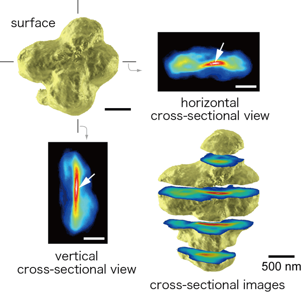 Three-dimensional image reconstructed of human chromosomes.
|
For more information, please contact: Dr. Kazuhiro Maeshima (RIKEN) |
 ,
,- Previous Article
- Clarification of Structure of Gibberellin Receptor - Spark for "Second Green Revolution" Enabling Complete Regulation of Plant Growth (Press Release)
- Current article
- Development of a New Type of X-Ray Microscopy for Three-Dimensional Observation of Cell Interior - Success in X-ray CT Scanning of Human Chromosomes for the First Time in the World (Press Release)