Irregular Packaging of DNA as Blueprint of Life Rather Than Conventionally Proposed Regular Packaging (Press Release)
- Release Date
- 18 Feb, 2012
- BL29XU (RIKEN Coherent X-ray Optics)
- BL45XU (RIKEN Structural Biology I)
Research Organization of Information and Systems
National Institute of Genetics
Key research achievements
• Finding that human genome DNA with a total length of 2 m is irregularly packaged in cell chromosomes
• Acquiring a clue for clarifying the mechanisms of packaging, search for, and readout of genetic information
Mitotic chromosomes (hereafter, chromosomes) are the bundles into which DNA*1 is compacted at the time of cell division (Fig. 1). Biology textbooks give an illustration of the following hierarchical structure (building-block structure); thin strands of DNA are wrapped around histone proteins to form nucleosomes, and the nucleosome fibers are regularly bundled to form chromatin fibers (Fig. 2). This established theory was proposed in the late 1970's and was hardly questioned by scientists. In this study, a research group led by Kazuhiro Maeshima (Professor) of the National Institute of Genetics has examined the structure of human chromosomes in detail using intense X-rays from a RIKEN beamline at SPring-8,*2 and obtained a finding that might overturn the conventional theory. They found that chromatin fibers were not regularly but irregularly bundled in chromosomes. The irregularly bundled structure indicates that cells produce chromosomes efficiently with minimum energy. The results of this study were published in the European Molecular Biology Organization (EMBO) Journal on 17 February 2012. Publication: |
<<Figures>>
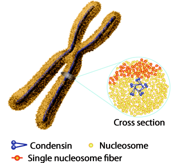
Fig. 1 Nucleosome fibers (red lines; see also Fig. 2) are irregularly packaged in chromosomes. Proteins called condensins (blue) and type II topoisomerases*3 exist and function as axes in chromosomes (left). Condensins bundle the nucleosome fibers to form a loop near the cross-sectional center (right), and the loop is considered to be irregularly packaged towards the center.
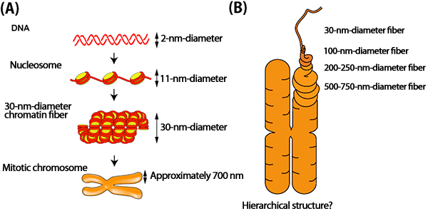
Fig. 2
(A) Thin DNA strands with a diameter of 2 nm (top column) wrap around histones to form nuclesome fibers with a diameter of approximately 11 nm (second column). The nulceosome fibers had long been considered to be regularly folded to form 30-nm-diameter chromatin fibers (third column).
(B) The conventionally proposed model explains that chromatin fibers form a regular helical hierarchical structure (building-block structure), i.e., they are helically wrapped to form 100-nm-diameter fibers, 200- to 250-nm-diameter fibers, then 500- to 750-nm-diameter fibers.
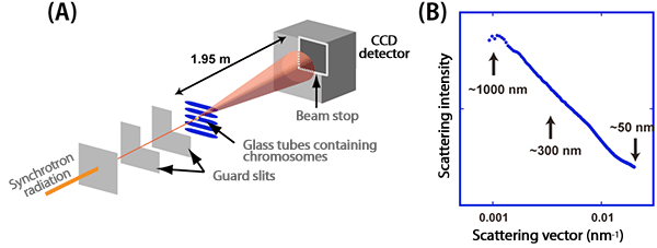
Fig. 3
(A) Chromosomes were exposed to synchrotron radiation generated from RIKEN Coherent X-ray Optics BL29XUL at SPring-8 to examine the scattering intensity in the range of the scattering vector of up to 1 µm, which is equivalent to the diameter of chromosomes.
(B) Detailed analysis revealed no scattering peaks corresponding to approximately 100 and 200-250 nm, which is expected on the basis of the conventional theory (Fig. 2).
<<Glossary>>
*1 Deoxyribonucleic acid (DNA)
DNA, the blueprint of life, has a structure in which two very thin strands are helically wrapped around a single axis. The two strands are bridged by base pair “ladders” as genetic codes. The diameter of the double helix is approximately 2 nm, and the total length of human DNA reaches 2 m. DNA is packaged into 46 bundles so as not to be cut or become tangled during cell division and to form chromosomes. These bundles of DNA are allotted to future cells called daughter cells after replication.
*2 SPring-8
SPring-8 is a facility that generates the world's highest-performance synchrotron radiation. It is located in Harima Science Garden City in Hyogo prefecture and is owned by RIKEN. The name SPring-8 is derived from Super Photon ring-8 GeV. Synchrotron radiation is an extremely powerful light that is obtained when the direction of electrons is bent in magnetic fields.
*3 Condensins and type II topoisomerase
Condensins, which consist of five subunits, and type II topoisomerase are large protein complexes that are essential for the formation of chromosomes. Both exist in chromosomes functioning as axes (Fig. 1). Condensins were discovered in 1997 by a research group led by Tatsuya Hirano, currently a chief scientist of RIKEN. Type II topoisomerase is an enzyme that untangles tangled strands of DNA, and their inhibitors are widely used as anticancer drugs.
|
For more information‚ please contact: |
- Current article
- Irregular Packaging of DNA as Blueprint of Life Rather Than Conventionally Proposed Regular Packaging (Press Release)