SPring-8, the large synchrotron radiation facility

Skip to content
» JAPANESE
Personal tools
 

Development of High-Resolution and Highly Reliable Imaging Method for Biological Samples -Twofold or greater improvement of resolution under conventional measurement conditions- (Press Release)

Release Date
29 Jan, 2015
  • SACLA BL3

RIKEN

A joint research group (RIKEN SPring-8 Center and Keio University) has developed a measurement and analysis method that can significantly improve the resolution and reliability of imaging of biological samples, such as cells, using coherent X-ray diffraction imaging (CXDI)*1, and demonstrated that resolution is improved by a factor of two or more by computer simulations. The work was carried out by Yuki Takayama (special postdoctoral researcher), Koji Yonekura (associate chief scientist) et al. of the Biostructural Mechanism Laboratory, RIKEN SPring-8 Center, and the group of Keio University.

CXDI is an imaging technique for observing the internal structure of samples of micrometer- to submicrometer-order size (from one-thousandth to ten-thousandth of a millimeter) that are extremely difficult or impossible to crystallize. This technique enables visualization at a resolution higher than that of optical microscopy without preparing slices of samples as done for electron microscopy. Recently, the technique has been applied to the structural analysis of biological samples such as cells. With the use of the X-ray free electron laser (XFEL)*2, an extremely intense coherent X-ray light source, imaging at a resolution of 30–60 nm (one nanometer is one-billionth of a meter) has become possible.

However, further improvement of the resolution of CXDI has been impossible because only weak signals can be obtained from biological samples with a low X-ray diffraction signals. In addition, diffraction patterns cannot be obtained in regions within a small diffraction angle range owing to experimental restrictions, and the conventional calculation algorithm used for image reconstruction cannot reconstruct the images in some cases.

The joint research group developed a new measurement and analysis method that can simultaneously image a biological sample and many gold particles with a high X-ray diffraction performance to address the above problems. By interference between the diffraction X-rays from the biological sample and gold particles, the diffraction signals from the biological sample are enhanced to a detectable level. In addition, a highly reliable image of the sample is reproduced by applying the positional information of gold particles, obtained from a diffraction pattern with a relatively intense signal from gold particles by the Patterson superposition algorithm, which was originally developing for solving the phase problem in X-ray protein crystallography. Computer simulations using SACLA*3, RIKEN’s XFEL facility, demonstrated that the resolution was improved by a factor of two or more and that low-contrast structures such as flagellar filaments at the periphery of the sample were clearly recovered. If this technique is practically applied, cells and functional materials will be more finely visualized, contributing to the clarification of their functions and principles.

This study was supported by RIKEN’s Program for Special Postdoctoral Researchers, a Grant-in-Aid for Research Activity Start-Up from the Japan Society for the Promotion of Science (JSPS), RIKEN’s Strategic Research Program, and the X-ray Free Electron Laser Priority Strategy Program. The achievements of this study were published online in the British scientific journal Scientific Reports on 28 January 2015.

*Joint research group
RIKEN SPring-8 Center (RSC)
Biostructural Mechanism Laboratory, Photon Science Research Division
   Yuki Takayama (postdoctoral researcher)
   Koji Yonekura (associate chief scientist)
Bio-specimen Platform Group, Advanced Photon Technology Division
   Saori Maki-Yonekura (research scientist)
Department of Physics, Faculty of Science and Technology, Keio University
   Masayoshi Nakasako (professor, also a visiting senior scientist of SR Life Science Instrumentation Unit, RSC)
   Tomotaka Oroguchi (assistant professor, also a visiting research scientist of SR Life Science Instrumentation Unit, RSC)

Publication:
Scientific Reports
Title:"Signal-enhancement and Patterson-search phasing for high-spatial-resolution coherent X-ray diffraction imaging of biological objects"
Authors:  Yuki Takayama, Saori Maki-Yonekura, Tomotaka Oroguchi, Masayoshi Nakasako, Koji Yonekura
doi: 10.1038/srep08074


<<Figures>>


Fig. 1	CXDI experiment using XFEL as light source
Fig.1   CXDI experiment using XFEL as light source

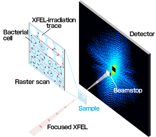
Sample particles dispersed on thin carbon film were irradiated with XFEL pulses to record the diffraction patterns using a two-dimensional detector. In the XFEL irradiation area, X-ray signals are explosively scattered after diffraction, and diffraction patterns are collected while moving the sample. A region within an extremely small angle in the diffraction patterns cannot be observed because of a beamstop that protects the detector from intense X-ray pulses.


Fig. 2	Test model for our new method and diffraction patterns calculated by computer simulations
Fig.2   Test model for our new method and diffraction patterns calculated by computer simulations

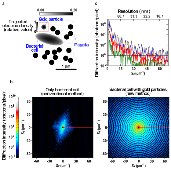
With the new method, a sample bacterium with many dispersed gold particles (a) is prepared and irradiated with XFEL pulses. In the diffraction pattern obtained from this sample (b, right), a diffraction pattern with a high intensity is observed for a diffraction angle more than twofold larger than that in the case of the diffraction pattern with the bacterium alone (b, left). The resolution is approximately 14 nm at the periphery. (c) Intensity profile along the red horizontal line in the diffraction pattern. The curves are as follows: blue solid line, the entire sample; yellow dotted line, the gold particles; green solid line, the bacterium; and the red solid line, the interference signal (absolute value) between the two. Because of the interference, the diffraction signal from the bacterial cell was confirmed to be enhanced by one order of magnitude. The signal originating from the gold particles contributes approximately 80% to the total signal and mostly overlaps the plot for the entire sample. Computer simulations are based on CXDI experiments using SACLA carried out by the joint research group.


Fig. 3	Comparison of density maps reconstructed by conventional and new methods
Fig.3   Comparison of density maps reconstructed by conventional and new methods

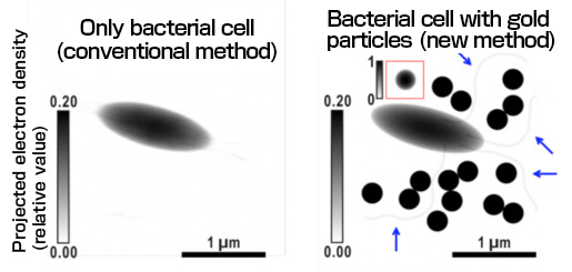
The density map reconstructed by the new method (right) accurately reproduces even flagella (indicated by arrows) inside and outside the bacterial cell compared with that reconstructed from the same bacterial image by the conventional method (left). The resolution was calculated to be 13 nm, which is higher than that of the conventional method (29 nm) by more than a factor. Projected electron density (relative value)


<<Glossary>>
*1  Coherent X-ray diffraction imaging (CXDI)

Imaging method utilizing the scattering phenomenon of X-rays. When a sample is irradiated with spatially coherent X-rays, a characteristic pattern (X-ray diffraction pattern) that reflects the sample structure is observed. The sample structure can be reconstructed from the diffraction pattern, when the pattern is recorded at a finer interval than a given value on the detector.

*2  X-ray free electron laser (XFEL)
Pulse laser in an X-ray region realized by the recent development of accelerator technology. Unlike conventional lasers using semiconductors and gases as the oscillation medium, XFEL uses electron beams that travel at a high speed in vacuum and has theoretically no restrictions on the wavelength used.

*3  SACLA
Japan’s first XFEL facility constructed jointly by RIKEN and Japan Synchrotron Radiation Research Institute. As one of the five national key technologies, the technology center was constructed and developed in a five-year project starting from FY 2006. It was completed in March 2011 and named SACLA taken from the initial letters of SPring-8 Angstrom Compact Free Electron LAser. The first successful generation of an X-ray laser occured in June 2011. Public operations began in March 2012. SACLA generates highly intense and highly coherent X-rays required for CXDI as very short (fs) pulses.


For more information, please contact:
Associate chief scientist Koji Yonekura (RIKEN SPring-8 Center (RSC))
Postdoctoral researcher Yuki Takayama (RIKEN SPring-8 Center (RSC))
TEL:+81-(0)791-58-2837 FAX:+81-(0)791-58-1844
E-mail:mail1

Previous Article
Synchrony of beating between implanted iPS cells and the host heart confirmed at a molecular level -a significant advance for cardiac regenerative medicine using iPS cells- (Press Release)
Current article
Development of High-Resolution and Highly Reliable Imaging Method for Biological Samples -Twofold or greater improvement of resolution under conventional measurement conditions- (Press Release)
Next Article
Clarification of Crystal Structure of Protein That “Removes” Causative Substance of Alzheimer’s Disease (Press Release)