Secondary structural analysis of Lewy bodies in the brain of Parkinson’s disease patients, a world first (Press Release)
- Release Date
- 01 Dec, 2015
- BL43IR (Infrared Materials Science)
2015-12-1
|
A group of researchers led by Katsuya ARAKI (Clinical Fellow) and Hideki MOCHIZUKI (Professor) at Osaka University Graduate School of Medicine, in cooperation with Dr. Naoto YAGI (Japan Synchrotron Radiation Research Institute (JASRI/SPring-8)), succeeded in elucidating the secondary structure of Lewy bodies (LBs) in the brain of Parkinson’s disease (PD) patients for the first time with synchrotron Fourier transform infrared micro-spectroscopy (FTIRM). LBs had been considered to be a key element of pathogenesis for PD. Although structural analysis for LBs with an electron microscope had been made, it had no secondary structural information of the protein, which is important for the development of drugs. In recent years, many researchers have focused on the new treatment to inhibit the formation of abnormal protein aggregates, which can delay the onset and progression of PD. This research result and method may provide important clues to the development of epoch-making treatment for PD. Abstract |
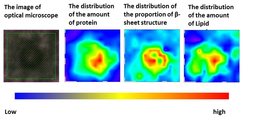
The central region (core) contains a large amount of proteins (second from left) and lipids (fourth from left). In contrast, the content of the β-sheet structure (third from left) is higher in the peripheral region (halo) than in the core.
To learn more about this research, please view the full research report entitled "Synchrotron FTIR micro-spectroscopy for structural analysis of Lewy bodies in the brain of Parkinson’s disease patients" at this page of the Scientific Reports website.
|
For more information, please contact: |

- Previous Article
- Grain boundary sliding as the major flow mechanism of Earth’s mantle (Press Release)
- Current article
- Secondary structural analysis of Lewy bodies in the brain of Parkinson’s disease patients, a world first (Press Release)