Clarification of Dynamic Structural Change of Calcium Pump
Growing attention paid to ion pump
Various ions are involved in the biological activities of living organisms. Ca2+ ions induce muscle movements and are one of the ions that are vital to living organisms (Fig. 1). Ca2+ ions are stored in the sarcoplasmic reticulum present around myofibrils. A muscle contracts when Ca2+ ions in the sarcoplasmic reticulum are released into muscle cells. In contrast, Ca2+ ions need to be pumped back into the sarcoplasmic reticulum to relax the muscle cells. A membrane protein called a calcium pump is responsible for the pumping back of Ca2+ ions.
To pump back a substance, the processes of grabbing and releasing are involved. Structural changes of the calcium pump corresponding to grabbing and releasing occur in the calcium pump during the pumping back of Ca2+ ions. Chikashi Toyoshima, a professor of the Institute of Molecular and Cellular Biosciences of The University of Tokyo, discovered this structural change ahead of other research groups worldwide. After 20 years of research, he found the structure of the calcium pump under nine different states.
Results of the analysis on the calcium pump structures clarified thus far were published in the British scientific journal, Nature, in 2000, 2002, and 2004. For this series of achievements, Professor Toyoshima was awarded The Asahi Prize in 2009, which is given to individuals or groups for their excellent achievements in the fields of academics, art, and others.
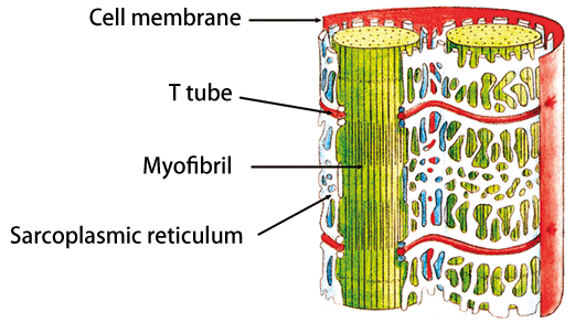
Fig. 1 Mechanism of muscle contraction
Muscles contract when Ca2+ ions are released from the sarcoplasmic reticulum around myofibrils; in contrast, muscles relax when Ca2+ ions are pumped back into the sarcoplasmic reticulum. The calcium pumps embedded in the sarcoplasmic reticulum membrane are responsible for the pumping back process.
Sample obtained by chance
The first trigger that started this research dates back 20 years when Professor Toyoshima studied in the UK as a young researcher. At that time, he analyzed the steric structure of a channel protein*1 using an electron microscope and developed a technique of analyzing the structure of tubular crystals. When he was searching for proteins that can be used to verify the general applicability of his technique, a researcher in a neighboring laboratory synthesized a tubular crystal of a calcium pump. Thus, the analysis of the steric structure of the calcium pump began because the calcium pump is a commonly available protein that can form tubular crystals. As the research progressed, he changed the analysis method from electron microscopy to X-ray crystallography in order to examine the steric structure of the proteins in more detail.
Out-of-the-box crystallization technique
For the analysis of the steric structure of proteins, crystals with a size and a uniformity (precision in terms of alignment) that are suitable for the analytical technique used should be prepared. However, it was impossible to synthesize the crystals of a calcium pump, a membrane protein embedded in a biological membrane, by conventional crystallization methods because the calcium pump cannot maintain its structure once removed from the biological membrane. At that time, it was considered that the crystallization of proteins embedded in a membrane is impossible.
Nonetheless, Professor Toyoshima succeeded in crystallizing the calcium pump embedded in a membrane. First, calcium pumps were dissolved from the biological membrane and purified to remove nontarget proteins. Then, the obtained proteins were crystallized by gradually changing the conditions of the solution. During this process, he added some lipids. As a result, crystals in which calcium pumps are embedded in a double lipid membrane were obtained. At first, the crystal is very thin, consisting of 10 layers of double lipid membrane. By changing the crystallization conditions, the thickness of the crystal was increased to a level that allows the crystal to be analyzed using intense X-rays at Spring-8 (Fig. 2).
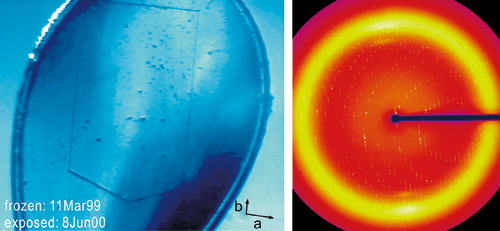
Fig. 2
Planar crystal of calcium pump (left) that is raised by a nylon loop and rapidly frozen (width, 300 μm; thickness, 20 μm). X-ray diffraction pattern (right) of planar crystal of calcium pump observed using BL41XU beamline.
Continuing interest
The structure of a calcium pump to which two Ca2+ ions are bound was reported in 2000 as the first achievement of this research (upper left of Fig. 3). The structure was determined using two beamlines, BL41XU and BL44B2, of SPring-8. As shown in the figure, the pump has two Ca2+ ions. “I could not have observed this structure without using the beamlines at SPring-8,” says Professor Toyoshima. With this achievement as a start, he decided to explore intensely how the calcium pump behaves.
The second structure reported in 2002 was the structure of a calcium pump that has released two Ca2+ ions (lower left of Fig. 3). Compared with the first structure, three domains*2 (A, N, P) come closer, and the fourth helix*3 (M4 helix) falls like a piston of a mechanical pump to push out the Ca2+ ions.
In 2004, Professor Toyoshima clarified the steric structure of the calcium pump, to which ATP*4, an energy source used to move muscles, is bound. As shown in the structure, Ca2+ ions were trapped by ATP (upper right of Fig. 3). In the same year, two more steric structures of the intermediate state were reported, further advancing the research in this field. The intermediate state is an instantaneous state during the reaction cycle related to Ca2+ ion transport. Therefore, a technique to stabilize the intermediate state is necessary for the crystallization of calcium pumps.
“Nobody had imagined that two domains rotate by 110° or a helix is dislocated by 10 Å (an angstrom is one-ten billionth of a meter). The structural change is more dynamic than we expected, which is intriguing and I want to examine this in more detail,” says Professor Toyoshima. This enthusiasm is his energy source for tackling the next difficult themes.
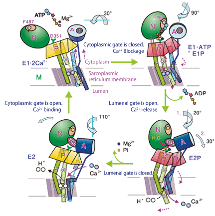
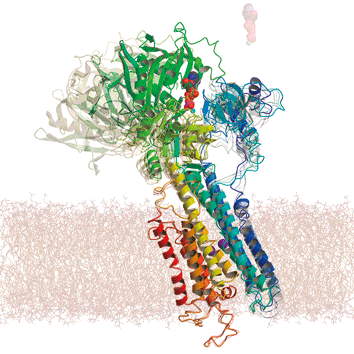
Fig. 3 Schematic of four basic states of calcium pump
Professor Toyoshima clarified over a long period the structural change shown in the figure.
Lower left: In the absence of two Ca2+ ions, three domains (A, N, P) on the cytoplasmic side come closer.
Upper left: When two Ca2+ ions are bound, the M5 helix straightens, and the three domains in the cytoplasm are well separated.
Upper right: The structure when ATP is bound, which was reported in 2004. Two Ca2+ ions are trapped.
Lower right: The M4 helix falls like a piston of a mechanical pump, releasing two Ca2+ ions. As a result, the Ca2+ ions are pumped back into the lumen of the sarcoplasmic reticulum.
Future direction of research
“What is important is not to merely clarify the structure, but to understand what you can learn from the structure. My superior whom I respect told me, ‘Nature is more complicated than physics researchers imagine.’ Maybe my senior said so because simplification is frequently carried out in physics to pursue a principle, and I really agree with him,” says Professor Toyoshima. From such a feeling, Professor Toyoshima, who majored in physics, can systematically continue his research to clarify the functions of the calcium pump.
A research group in Denmark also started this unique study around 2004, and there is stiff competition between the related research groups. A research institute that specializes in ion pump research was established in Denmark to expand the research field not only in terms of its structure but also in the application of drugs and medicines. The research on the calcium pump has been active because it is hoped that this research will be useful for the treatment of myocardial infarction and cancer. Under such circumstances, Professor Toyoshima remains calm because he thinks that such frontier research cannot be carried out by anyone without sufficient preparation. He is steadily preparing to explore the two intermediate states that trigger the functioning of the calcium pump.
Professor Toyoshima decided that the next research target is the sodium-potassium pump and has already started the research. He has already begun making some progress and this interviewer is looking forward to finding out how he will interpret the structure and function of the pump.
Glossary
*1 Channel protein
A channel protein transports only specific ions in a biological membrane, and scarcely transfers substances. An ion pump pumps ions from the low-concentration side to the high-concentration side using ATP energy. In contrast, a channel serves as a gate that allows the flow of ions from the high-concentration side to the low-concentration side.
*2 Domain
A domain is a part of a protein structure and functions collectively.
*3 Helix
A helix is a part of a protein structure and forms a spiral like a spring.
*4 ATP
ATP is an abbreviation of adenosine triphosphate. Three phosphoric acids are bonded to an adenosine. Energy is released when the bonds of the phosphoric molecules are broken.
Make what is necessary by myself
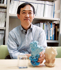 |
| Professor Toyoshima with handmade models |
Professor Toyoshima likes to make things. He brought handmade models of a calcium pump and a calcium channel from his laboratory to show to the interviewer. He makes gadgets and develops computer programs when he thinks they will be useful in his experiment. Thus, he increases the efficiency of the experiment and makes the experimental procedure easier. His idea was also applied to a beamline of SPring-8. He believes that making something by himself is important to start something new, showing the spirit of a developer when he was involved in the technological development of an electron microscope.
Behind his significant research achievements is careful attention to experimental details.
 |
Schematic of structure: Structural change of calcium pump One step of the reaction cycle of a calcium pump. This is a state when ATP is bound and Ca2+ ions are trapped in the membrane. The intermediate structure shown in gray was determined by calculation |
Interview and original text by Akiko Ikeda (Sci-Tech Communications Incorporated)
This article was written following an interview with Professor Chikashi Toyoshima at the Institute of Molecular and Cellular Biosciences of the University of Tokyo.