Topic 6:Imaging of Newborn Rabbit Lungs
Elucidation of “How Newborns Start Breathing?”
A fetus does not require oxygen exchange via the pulmonary system; consequently, the lungs are filled with “lung water” until birth. Immediately after birth, however, the lung water must be removed so that air can enter the lungs, thereby the newborn can begin breathing in a normal way. If this process does not go well, a newborn cannot breathe, and it will die. To avoid such an event, we need to understand the mechanisms by which a newborn's lungs begin breathing. However, because there have heretofore been no practical methods for observing the process of lung water being replaced with air, the details have remained unclear - but that is where SPring-8 comes in. The process of the initiation of breathing has now been observed, yielding many new discoveries about the lungs.
Image Analysis Techniques using X-ray Refraction
While in the womb, a fetus gets oxygen from the mother's blood through the placenta. The placenta acts as a filter, providing oxygen to a fetus through the umbilical cord. Upon birth, the lungs are filled with air and breathing begins. Most newborns manage this without difficulty, but in fact it is not an easy task at all. Neonatal asphyxia, in which a newborn cannot begin breathing on its own and therefore requires resuscitation, occurs in ~10% of deliveries.
How does a newborn inhale air into the lungs to begin breathing? The amount of lung water that a newborn expels from the mouth is small compared to the volume of the lungs. Thus, it is unclear how lung water is eliminated. It has been speculated that lung water gradually decreases over time, and is replaced with air by some unknown mechanism. One hypothesis was that osmotic gradient created by epithelial Na channel gradually moves water across the wall of alveolus. In 2003, a collaborative research group consisting of researchers from JASRI and Monash University in Melbourne, Australia initiated research with the aim of unraveling this mystery.
At SPring-8, researchers from JASRI have been playing a central role in developing the novel technique of refraction contrast imaging. This is a method for obtaining density distribution images with enhanced edge contrast by exploiting the characteristics of X-rays. In refraction contrast imaging, X-rays are superimposed at the edge of an object according to the slight differences in the refractive index of the object's materials (Fig. 1). The diffraction angle is extremely small (0.1 mm displacement at a distance of 10 m from the object) but the high-resolution image detector developed at JASRI can distinguish such a small angle. Using this technique, we can obtain higher-resolution images than those obtained with existing radiographic visualization techniques (absorption contrast imaging) such as medical X-ray imaging, which exploits the differences between an object's absorption and penetration by X-rays.
Collimated (i.e., perfectly parallel) X-rays are essential for detecting images based on the extremely small differences in the refractive index. Generation of collimated X-rays is among the special technologies that are available at SPring-8.
The energy of X-ray employed also differs between refraction contrast imaging and absorption contrast imaging. The higher the X-ray energy, the higher the penetration and the lower the absorption. When high-energy X-rays are used, satisfactory data will not be obtained unless there are sufficient differences in absorption in soft tissues. However, even in such a situation, differences in the refractive index will be useful. On the other hand, when low-energy X-rays are used, information about tissues in front of and behind a hard tissue cannot be obtained. By using refracted waves of high-energy X-rays, the structures of multi-layered hard and soft tissues can be observed.
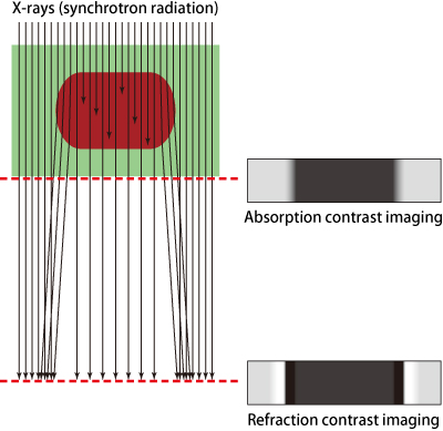
When X-rays (arrow) enter an object (brown), X-rays are partially absorbed by the object and refracted at the surface as well. Refraction changes the direction of the X-rays. X-rays recorded immediately behind the object yield images with contrast caused primarily by absorption, but with only small effects from refraction (absorption contrast imaging). On the other hand, when X-rays are recorded at a distant location, both refracted and transmitted X-rays overlap to generate bright regions. This overlap enhances the edge of the object, allowing high-contrast images to be obtained (refraction contrast imaging).
Air Compresses Lung Water - Unexpected Phenomenon
The Monash University research team wrestled with the observation of the lungs of newborn rabbits by applying the refraction contrast imaging that SPring-8 has developed over the last 10 years.
At the 220-m-long Medical and Imaging I Beamline (BL20B2), samples were placed at 210 m from the deflection electromagnets that produce X-rays, and a detector was installed 2 m behind the samples. Since it was difficult to control the delivery timing, fetuses were taken out by cesarean section.
However, some special manipulation is required to observe temporal changes, since newborns begin breathing almost immediately after birth. Newborns begin breathing after the amnion, which is a thin membrane covering the fetus, is removed. To control the timing of breath initiation, rabbit fetuses were taken out from the womb and placed in warm water containers (37°C) without removing the amnion. They were immobilized in the containers, and X-rays were controlled so that they ran parallel to the fetal chest. Next, the amnion was removed, and irradiation was initiated in order to start the observation.
Irradiated X-rays entered the detector after experiencing a slight refraction. X-rays that penetrated the newborn's body also entered the detector. Data were continuously recorded every 0.8 sec and processed by computer. Realistic lung images were displayed on a display monitor (Fig. 2). How is lung water replaced with air? These images of newborn rabbits revealed an unexpected phenomenon. When newborns begin breathing, air simply enters and fills the lungs upon each breath.
“It looks like air enters the lungs upon breathing and compresses the water inside the lungs. This unexpected phenomenon suggests that inspiration plays an important role in removing lung water,” explained Dr. Naoto Yagi (Chief Scientist, JASRI). Dr. Yagi is an expert in X-ray image analysis who has been participating in the development of refraction contrast imaging. He has played a central role in the collaboration with Monash University.
The respiratory organs cooperatively work to push lung water deep into the lungs; therefore, backflow upon exhalation was much smaller than the advance associated with inhalation. It is speculated that upon breathing thorax expands and lung water is sucked into tissues through the walls of peripheral organs such as the alveoli, and is gradually processed via the lymphatic and blood vessels.
This research group has succeeded for the first time in elucidating the mechanisms of breath initiation at birth, by visualizing the process of air entering into the lungs of newborn mammals. This research achievement was published in FASEB Journal (2007), a major journal for biomedical researches in the US.
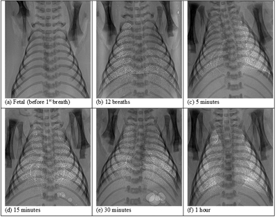
(a) Prior to initial breathing. The lungs are filled with lung water and airways are not observed.
(b-f) After initiation of breathing (numbers indicate elapsed time after the initiation of breathing), the tracheae and bronchi are visible. Additionally, increasing number of white spots show the delivery of oxygen into the peripheral parts of the lungs. At 30 and 60 min, the edges of the lungs and the existence of the diaphragm can be clearly identified.
Mechanical Ventilation that Compresses Lung Water is Effective
If lung water remains in the lungs after birth, the newborn will go into respiratory distress. To avoid this, mechanical ventilation is required for premature infants or newborns with respiratory disorder. The same mechanical ventilation used for adults has been applied in both premature infants and newborns.
However, now that the research conducted by Dr. Yagi and colleagues including researchers of Monash University revealed that newborns push lung water deep into the lungs by breathing in, we must reconsider how to use ventilation in underdeveloped lungs of premature infants without causing injury. Therefore, procedures for mechanical ventilation of newborns must be significantly revised. Lung water should be pushed by air using mechanical ventilation. A limited number of hospitals have applied such mechanical ventilation, but this treatment has not been widespread. After this treatment demonstrated the efficacy, however, increasing numbers of hospitals in Australia and the Netherlands have been adopting this treatment method.
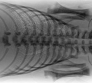
Lying sideways, as shown in this figure, often causes the non-uniform entry of air across the upper and lower parts of the lungs. A larger amount of air, indicated by white spots, enters the upper part; however, air enters only the major bronchi in the lower part.
References
1. S. B. Hooper, M. J. Kitchen, M. J. Wallace, N. Yagi, K. Uesugi, M. J. Morgan, C. Hall, K. K. W. Siu, I. M. Williams, M. Siew, S. C. Irvine, K. Pavlov and R. A. Lewis; FASEB J., 21, 3329 (2007)
2. A. B. te Pas, M. Siew, M. J. Wallace, M. J. Kitchen, A. Fouras, R. A. Lewis, N. Yagi, K. Uesugi, S. Donath, P. G. Davis, C. J. Morley and S. B. Hooper; Pediatric Research, 65, 537 (2009)